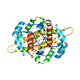
4PJ1
 
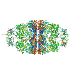 | | Crystal structure of the human mitochondrial chaperonin symmetrical 'football' complex | | 分子名称: | 10 kDa heat shock protein, mitochondrial, 60 kDa heat shock protein, ... | | 著者 | Frolow, F, Azem, A, Nisemblat, S. | | 登録日 | 2014-05-10 | | 公開日 | 2015-04-29 | | 最終更新日 | 2023-09-27 | | 実験手法 | X-RAY DIFFRACTION (3.15 Å) | | 主引用文献 | Crystal structure of the human mitochondrial chaperonin symmetrical football complex.
Proc.Natl.Acad.Sci.USA, 112, 2015
|
|
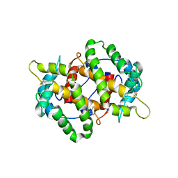
6MRC
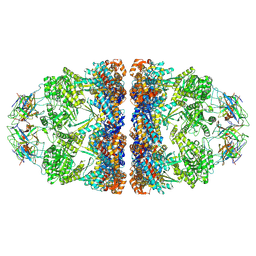 | | ADP-bound human mitochondrial Hsp60-Hsp10 football complex | | 分子名称: | 10 kDa heat shock protein, mitochondrial, 60 kDa heat shock protein, ... | | 著者 | Gomez-Llorente, Y, Jebara, F, Patra, M, Malik, R, Nissemblat, S, Azem, A, Hirsch, J.A, Ubarretxena-Belandia, I. | | 登録日 | 2018-10-12 | | 公開日 | 2020-04-15 | | 最終更新日 | 2024-03-13 | | 実験手法 | ELECTRON MICROSCOPY (3.08 Å) | | 主引用文献 | Structural basis for active single and double ring complexes in human mitochondrial Hsp60-Hsp10 chaperonin.
Nat Commun, 11, 2020
|
|
6MRD
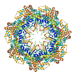 | | ADP-bound human mitochondrial Hsp60-Hsp10 half-football complex | | 分子名称: | 10 kDa heat shock protein, mitochondrial, 60 kDa heat shock protein, ... | | 著者 | Gomez-Llorente, Y, Jebara, F, Patra, M, Malik, R, Nissemblat, S, Azem, A, Hirsch, J.A, Ubarretxena-Belandia, I. | | 登録日 | 2018-10-12 | | 公開日 | 2020-04-15 | | 最終更新日 | 2024-03-13 | | 実験手法 | ELECTRON MICROSCOPY (3.82 Å) | | 主引用文献 | Structural basis for active single and double ring complexes in human mitochondrial Hsp60-Hsp10 chaperonin.
Nat Commun, 11, 2020
|
|
6HT7
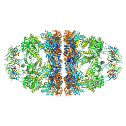 | | Crystal structure of the WT human mitochondrial chaperonin (ADP:BeF3)14 complex | | 分子名称: | 10 kDa heat shock protein, mitochondrial, 60 kDa heat shock protein, ... | | 著者 | Jebara, F, Patra, M, Azem, A, Hirsch, J. | | 登録日 | 2018-10-03 | | 公開日 | 2020-04-29 | | 最終更新日 | 2024-06-19 | | 実験手法 | X-RAY DIFFRACTION (3.7 Å) | | 主引用文献 | Crystal structure of the WT human mitochondrial football Hsp60-Hsp10(ADPBeFx)14 complex
Nat Commun, 2020
|
|
4IJ7
 | | Crystal structure of Odorant Binding Protein 48 from Anopheles gambiae (AgamOBP48) with PEG | | 分子名称: | 2,5,8,11,14,17,20,23,26,29,32,35,38,41,44,47,50,53,56,59,62,65,68,71,74,77,80-HEPTACOSAOXADOOCTACONTAN-82-OL, Odorant binding protein-8, SODIUM ION | | 著者 | Zographos, S.E, Tsitsanou, K.E, Drakou, C.E. | | 登録日 | 2012-12-21 | | 公開日 | 2013-10-16 | | 最終更新日 | 2023-09-20 | | 実験手法 | X-RAY DIFFRACTION (2.25 Å) | | 主引用文献 | Crystal and Solution Studies of the "Plus-C" Odorant-binding Protein 48 from Anopheles gambiae: CONTROL OF BINDING SPECIFICITY THROUGH THREE-DIMENSIONAL DOMAIN SWAPPING.
J.Biol.Chem., 288, 2013
|
|
4KYN
 | |


