8CRA
 
 | |
6CIG
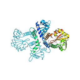
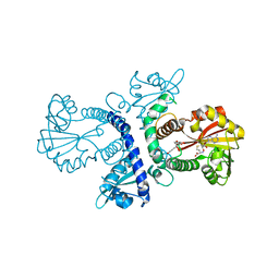
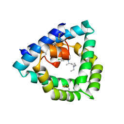
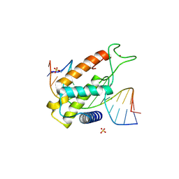
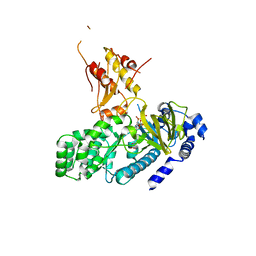
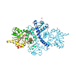 | | CRYSTAL STRUCTURE ANALYSIS OF SELENOMETHIONINE SUBSTITUTED ISOFLAVONE O-METHYLTRANSFERASE | | 分子名称: | GLYCEROL, Isoflavone-7-O-methyltransferase 8, N-(TRIS(HYDROXYMETHYL)METHYL)-3-AMINOPROPANESULFONIC ACID, ... | | 著者 | Zubieta, C, Dixon, R.A, Shabalin, I.G, Kowiel, M, Porebski, P.J, Noel, J.P. | | 登録日 | 2018-02-23 | | 公開日 | 2018-03-07 | | 最終更新日 | 2022-03-23 | | 実験手法 | X-RAY DIFFRACTION (1.65 Å) | | 主引用文献 | Structures of two natural product methyltransferases reveal the basis for substrate specificity in plant O-methyltransferases.
Nat. Struct. Biol., 8, 2001
|
|
6SLR
 | |
2VDJ
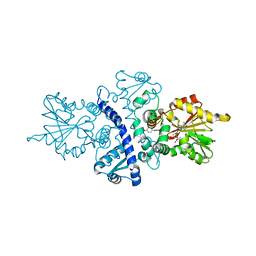
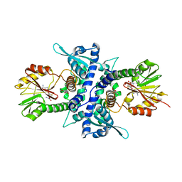
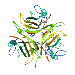
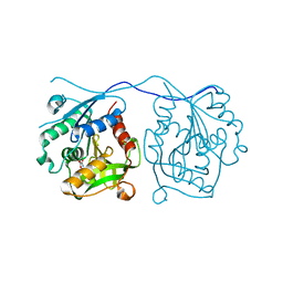 | | Crystal Structure of Homoserine O-acetyltransferase (metA) from Bacillus Cereus with Homoserine | | 分子名称: | HOMOSERINE O-SUCCINYLTRANSFERASE, L-HOMOSERINE, SULFATE ION | | 著者 | Zubieta, C, Arkus, K.A.J, Cahoon, R.E, Jez, J.M. | | 登録日 | 2007-10-10 | | 公開日 | 2008-01-22 | | 最終更新日 | 2023-12-13 | | 実験手法 | X-RAY DIFFRACTION (2 Å) | | 主引用文献 | A Single Amino Acid Change is Responsible for Evolution of Acyltransferase Specificity in Bacterial Methionine Biosynthesis.
J.Biol.Chem., 283, 2008
|
|
5LXU
 | |
1X9T
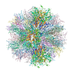
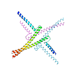
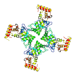
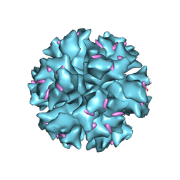 | | The crystal structure of human adenovirus 2 penton base in complex with an ad2 N-terminal fibre peptide | | 分子名称: | N-DODECYL-N,N-DIMETHYL-3-AMMONIO-1-PROPANESULFONATE, N-terminal peptide of Fiber protein, Penton protein | | 著者 | Zubieta, C, Schoehn, G, Chroboczek, J, Cusack, S. | | 登録日 | 2004-08-24 | | 公開日 | 2005-01-18 | | 最終更新日 | 2024-04-03 | | 実験手法 | X-RAY DIFFRACTION (3.5 Å) | | 主引用文献 | The structure of the human adenovirus 2 penton
Mol.Cell, 17, 2005
|
|
1X9P
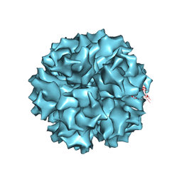 | | The crystal structure of human adenovirus 2 penton base | | 分子名称: | N-DODECYL-N,N-DIMETHYL-3-AMMONIO-1-PROPANESULFONATE, Penton protein, SULFATE ION | | 著者 | Zubieta, C, Schoehn, G, Chroboczek, J, Cusack, S. | | 登録日 | 2004-08-24 | | 公開日 | 2005-01-18 | | 最終更新日 | 2024-04-03 | | 実験手法 | X-RAY DIFFRACTION (3.3 Å) | | 主引用文献 | The structure of the human adenovirus 2 penton
Mol.Cell, 17, 2005
|
|
1FPQ
 | |
1FP2
 | |
1FP1
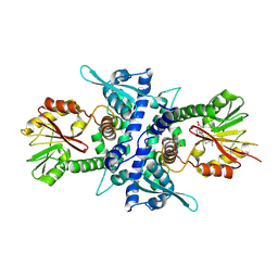
 | | CRYSTAL STRUCTURE ANALYSIS OF CHALCONE O-METHYLTRANSFERASE | | 分子名称: | 2',4,4'-TRIHYDROXYCHALCONE, ISOLIQUIRITIGENIN 2'-O-METHYLTRANSFERASE, S-ADENOSYL-L-HOMOCYSTEINE | | 著者 | Zubieta, C, Dixon, R.A, Noel, J.P. | | 登録日 | 2000-08-29 | | 公開日 | 2001-03-07 | | 最終更新日 | 2024-02-07 | | 実験手法 | X-RAY DIFFRACTION (1.82 Å) | | 主引用文献 | Structures of two natural product methyltransferases reveal the basis for substrate specificity in plant O-methyltransferases.
Nat.Struct.Biol., 8, 2001
|
|
1KYZ
 | | Crystal Structure Analysis of Caffeic acid/5-hydroxyferulic acid 3/5-O-methyltransferase Ferulic Acid Complex | | 分子名称: | 3-(4-HYDROXY-3-METHOXYPHENYL)-2-PROPENOIC ACID, Caffeic acid 3-O-methyltransferase, S-ADENOSYL-L-HOMOCYSTEINE | | 著者 | Zubieta, C, Kota, P, Ferrer, J.-L, Dixon, R.A, Noel, J.P. | | 登録日 | 2002-02-06 | | 公開日 | 2002-08-28 | | 最終更新日 | 2024-04-03 | | 実験手法 | X-RAY DIFFRACTION (2.2 Å) | | 主引用文献 | Structural basis for the modulation of lignin monomer methylation by caffeic acid/5-hydroxyferulic acid 3/5-O-methyltransferase.
Plant Cell, 14, 2002
|
|
1KYW
 | | Crystal Structure Analysis of Caffeic Acid/5-hydroxyferulic acid 3/5-O-methyltransferase in complex with 5-hydroxyconiferaldehyde | | 分子名称: | 5-(3,3-DIHYDROXYPROPENY)-3-METHOXY-BENZENE-1,2-DIOL, Caffeic acid 3-O-methyltransferase, S-ADENOSYL-L-HOMOCYSTEINE | | 著者 | Zubieta, C, Kota, P, Ferrer, J.-L, Dixon, R.A, Noel, J.P. | | 登録日 | 2002-02-06 | | 公開日 | 2002-08-28 | | 最終更新日 | 2024-04-03 | | 実験手法 | X-RAY DIFFRACTION (2.4 Å) | | 主引用文献 | Structural basis for the modulation of lignin monomer methylation by caffeic acid/5-hydroxyferulic acid 3/5-O-methyltransferase.
Plant Cell, 14, 2002
|
|
1M6E
 | | CRYSTAL STRUCTURE OF SALICYLIC ACID CARBOXYL METHYLTRANSFERASE (SAMT) | | 分子名称: | 2-HYDROXYBENZOIC ACID, LUTETIUM (III) ION, S-ADENOSYL-L-HOMOCYSTEINE, ... | | 著者 | Zubieta, C, Ross, J.R, Koscheski, P, Yang, Y, Pichersky, E, Noel, J.P. | | 登録日 | 2002-07-16 | | 公開日 | 2003-09-09 | | 最終更新日 | 2024-02-14 | | 実験手法 | X-RAY DIFFRACTION (3 Å) | | 主引用文献 | Structural Basis for Substrate Recognition in The Salicylic Acid Carboxyl Methyltransferase Family
Plant Cell, 15, 2003
|
|
2C6S
 | |
7NB0
 | |
6OMS
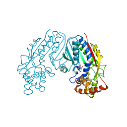
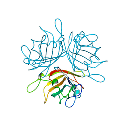
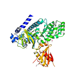
 | | Arabidopsis GH3.12 with Chorismate | | 分子名称: | (3R,4R)-3-[(1-carboxyethenyl)oxy]-4-hydroxycyclohexa-1,5-diene-1-carboxylic acid, 4-substituted benzoates-glutamate ligase GH3.12, ADENOSINE MONOPHOSPHATE | | 著者 | Zubieta, C, Westfall, C.S, Holland, C.K, Jez, J.M. | | 登録日 | 2019-04-19 | | 公開日 | 2019-10-09 | | 最終更新日 | 2023-10-11 | | 実験手法 | X-RAY DIFFRACTION (1.942 Å) | | 主引用文献 | Brassicaceae-specific Gretchen Hagen 3 acyl acid amido synthetases conjugate amino acids to chorismate, a precursor of aromatic amino acids and salicylic acid.
J.Biol.Chem., 294, 2019
|
|
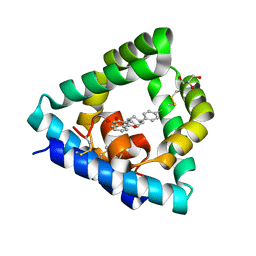
4V2O
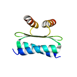
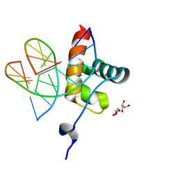
 | | Structure of saposin B in complex with chloroquine | | 分子名称: | N4-(7-CHLORO-QUINOLIN-4-YL)-N1,N1-DIETHYL-PENTANE-1,4-DIAMINE, SAPOSIN-B | | 著者 | Zubieta, C, Lai, X, Doyle, R.P. | | 登録日 | 2014-10-13 | | 公開日 | 2015-12-09 | | 最終更新日 | 2024-01-10 | | 実験手法 | X-RAY DIFFRACTION (2.13 Å) | | 主引用文献 | The Lysosomal Protein Saposin B Binds Chloroquine.
Chemmedchem, 11, 2016
|
|
4OX0
 | |
8P5Q
 | |
6QEC
 | |
6F6O
 | |
4L39
 | | Crystal structure of GH3.12 from Arabidopsis thaliana in complex with AMPCPP and salicylate | | 分子名称: | 2-HYDROXYBENZOIC ACID, 4-substituted benzoates-glutamate ligase GH3.12, DIPHOSPHOMETHYLPHOSPHONIC ACID ADENOSYL ESTER, ... | | 著者 | Zubieta, C, Jez, J.M, Brown, E, Marcellin, R, Kapp, U, Round, A, Westfall, C. | | 登録日 | 2013-06-05 | | 公開日 | 2013-10-02 | | 最終更新日 | 2023-09-20 | | 実験手法 | X-RAY DIFFRACTION (2.81 Å) | | 主引用文献 | Determination of the GH3.12 protein conformation through HPLC-integrated SAXS measurements combined with X-ray crystallography.
Acta Crystallogr.,Sect.D, 69, 2013
|
|
4LIY
 | |
3N6Q
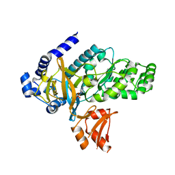
 | | Crystal structure of YghZ from E. coli | | 分子名称: | MAGNESIUM ION, YghZ aldo-keto reductase | | 著者 | Zubieta, C, Totir, M, Echols, N, May, A, Alber, T. | | 登録日 | 2010-05-26 | | 公開日 | 2011-06-15 | | 最終更新日 | 2023-09-06 | | 実験手法 | X-RAY DIFFRACTION (1.8 Å) | | 主引用文献 | Macro-to-Micro Structural Proteomics: Native Source Proteins for High-Throughput Crystallization.
Plos One, 7, 2012
|
|
4EQ4
 | | Crystal structure of seleno-methionine derivatized GH3.12 | | 分子名称: | 2-HYDROXYBENZOIC ACID, 4-substituted benzoates-glutamate ligase GH3.12, ADENOSINE MONOPHOSPHATE | | 著者 | Zubieta, C, Nanao, M, Jez, J, Westfall, C, Kapp, U. | | 登録日 | 2012-04-18 | | 公開日 | 2012-06-20 | | 最終更新日 | 2012-07-25 | | 実験手法 | X-RAY DIFFRACTION (2.074 Å) | | 主引用文献 | Structural basis for prereceptor modulation of plant hormones by GH3 proteins.
Science, 336, 2012
|
|


