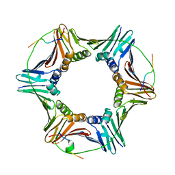
1CTN
 
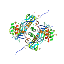
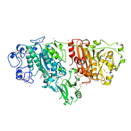
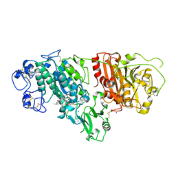
 | | CRYSTAL STRUCTURE OF A BACTERIAL CHITINASE AT 2.3 ANGSTROMS RESOLUTION | | 分子名称: | CHITINASE A | | 著者 | Perrakis, A, Tews, I, Dauter, Z, Wilson, K.S, Vorgias, C.E. | | 登録日 | 1994-10-10 | | 公開日 | 1995-02-07 | | 最終更新日 | 2024-10-30 | | 実験手法 | X-RAY DIFFRACTION (2.3 Å) | | 主引用文献 | Crystal structure of a bacterial chitinase at 2.3 A resolution.
Structure, 2, 1994
|
|
6QCG
 | |
8ARF
 | |
7P0K
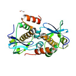
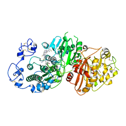 | | Crystal structure of Autotaxin (ENPP2) with 18F-labeled positron emission tomography ligand | | 分子名称: | 2-[[2-ethyl-6-[4-[2-[(3~{R})-3-fluoranylpyrrolidin-1-yl]-2-oxidanylidene-ethyl]piperazin-1-yl]imidazo[1,2-a]pyridin-3-yl]-methyl-amino]-4-(4-fluorophenyl)-2,3-dihydro-1,3-thiazole-5-carbonitrile, 2-acetamido-2-deoxy-beta-D-glucopyranose, CALCIUM ION, ... | | 著者 | Salgado-Polo, F, Shao, T, Xiao, Z, Van, R, Chen, J, Rong, J, Haider, A, Shao, Y, Josephson, L, Perrakis, A, Liang, S.H. | | 登録日 | 2021-06-29 | | 公開日 | 2022-07-13 | | 最終更新日 | 2024-10-16 | | 実験手法 | X-RAY DIFFRACTION (2.2 Å) | | 主引用文献 | Imaging Autotaxin In Vivo with 18 F-Labeled Positron Emission Tomography Ligands
J Med Chem, 64, 2021
|
|
6NVQ
 | |
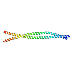
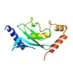
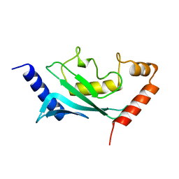
5C9N
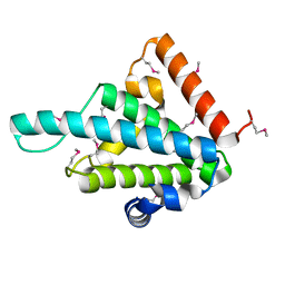
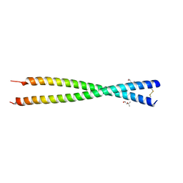 | | Crystal structure of GEMC1 coiled-coil domain | | 分子名称: | (4S)-2-METHYL-2,4-PENTANEDIOL, Geminin coiled-coil domain-containing protein 1 | | 著者 | Caillat, C, Perrakis, A. | | 登録日 | 2015-06-28 | | 公開日 | 2015-11-11 | | 最終更新日 | 2024-11-06 | | 実験手法 | X-RAY DIFFRACTION (2.2 Å) | | 主引用文献 | The structure of the GemC1 coiled coil and its interaction with the Geminin family of coiled-coil proteins.
Acta Crystallogr.,Sect.D, 71, 2015
|
|
2V0S
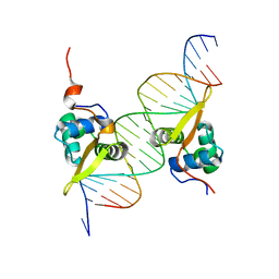
 | | crystal structure of a hairpin exchange variant (LR1) of the targeting LINE-1 retrotransposon endonuclease | | 分子名称: | GLYCEROL, LR1, MANGANESE (II) ION, ... | | 著者 | Repanas, K, Zingler, N, Layer, L.E, Schumann, G.G, Perrakis, A, Weichenrieder, O. | | 登録日 | 2007-05-17 | | 公開日 | 2007-07-17 | | 最終更新日 | 2023-12-13 | | 実験手法 | X-RAY DIFFRACTION (1.8 Å) | | 主引用文献 | Determinants for DNA Target Structure Selectivity of the Human Line-1 Retrotransposon Endonuclease.
Nucleic Acids Res., 35, 2007
|
|
2V0R
 | | crystal structure of a hairpin exchange variant (LTx) of the targeting LINE-1 retrotransposon endonuclease | | 分子名称: | LTX, SULFATE ION | | 著者 | Repanas, K, Zingler, N, Layer, L.E, Schumann, G.G, Perrakis, A, Weichenrieder, O. | | 登録日 | 2007-05-17 | | 公開日 | 2007-07-17 | | 最終更新日 | 2023-12-13 | | 実験手法 | X-RAY DIFFRACTION (2.3 Å) | | 主引用文献 | Determinants for DNA Target Structure Selectivity of the Human Line-1 Retrotransposon Endonuclease
Nucleic Acids Res., 35, 2007
|
|
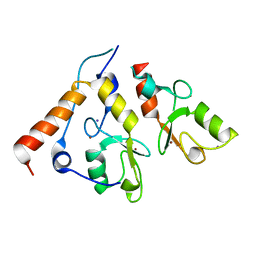
3KJY
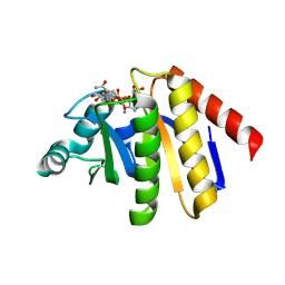
 | | Crystal structure of reduced HOMO SAPIENS CLIC3 | | 分子名称: | Chloride intracellular channel protein 3, SULFATE ION | | 著者 | Littler, D.R, Curmi, P.M.G, Breit, S.N, Perrakis, A. | | 登録日 | 2009-11-04 | | 公開日 | 2009-11-17 | | 最終更新日 | 2023-11-01 | | 実験手法 | X-RAY DIFFRACTION (1.95 Å) | | 主引用文献 | Structure of human CLIC3 at 2 A resolution
Proteins, 78, 2010
|
|
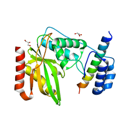
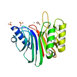
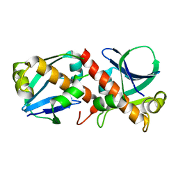
1U9B
 | | MURINE/HUMAN UBIQUITIN-CONJUGATING ENZYME UBC9 | | 分子名称: | UBIQUITIN-CONJUGATING ENZYME E9 | | 著者 | Tong, H, Hateboer, G, Perrakis, A, Bernards, R, Sixma, T.K. | | 登録日 | 1997-05-20 | | 公開日 | 1997-07-07 | | 最終更新日 | 2024-05-22 | | 実験手法 | X-RAY DIFFRACTION (2 Å) | | 主引用文献 | Crystal structure of murine/human Ubc9 provides insight into the variability of the ubiquitin-conjugating system.
J.Biol.Chem., 272, 1997
|
|
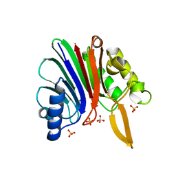
1U9A
 | | HUMAN UBIQUITIN-CONJUGATING ENZYME UBC9 | | 分子名称: | UBIQUITIN-CONJUGATING ENZYME | | 著者 | Tong, H, Hateboer, G, Perrakis, A, Bernards, R, Sixma, T.K. | | 登録日 | 1997-02-11 | | 公開日 | 1997-05-15 | | 最終更新日 | 2024-05-22 | | 実験手法 | X-RAY DIFFRACTION (2 Å) | | 主引用文献 | Crystal structure of murine/human Ubc9 provides insight into the variability of the ubiquitin-conjugating system.
J.Biol.Chem., 272, 1997
|
|
2CKL
 | | Ring1b-Bmi1 E3 catalytic domain structure | | 分子名称: | IODIDE ION, POLYCOMB GROUP RING FINGER PROTEIN 4, UBIQUITIN LIGASE PROTEIN RING2, ... | | 著者 | Buchwald, G, van der Stoop, P, Weichenrieder, O, Perrakis, A, van Lohuizen, M, Sixma, T.K. | | 登録日 | 2006-04-20 | | 公開日 | 2006-05-11 | | 最終更新日 | 2024-05-08 | | 実験手法 | X-RAY DIFFRACTION (2 Å) | | 主引用文献 | Structure and E3-Ligase Activity of the Ring-Ring Complex of Polycomb Proteins Bmi1 and Ring1B.
Embo J., 25, 2006
|
|
8A2F
 | | Crystal Structure of Ljunganvirus 1 2A protein | | 分子名称: | 2A protein | | 著者 | von Castelmur, E, Zhu, L, Wang, X, Fry, E, Ren, J, Perrakis, A, Stuart, D.I. | | 登録日 | 2022-06-03 | | 公開日 | 2023-06-14 | | 最終更新日 | 2024-02-07 | | 実験手法 | X-RAY DIFFRACTION (1.73 Å) | | 主引用文献 | Structural plasticity of 2A proteins in the Parechovirus family
To Be Published
|
|
8A2E
 | | Crystal Structure of Human Parechovirus 3 2A protein | | 分子名称: | 2A protein, GLYCEROL, SULFATE ION | | 著者 | von Castelmur, E, Zhu, L, wang, X, Fry, E, Ren, J, Perrakis, A, Stuart, D.I. | | 登録日 | 2022-06-03 | | 公開日 | 2023-06-14 | | 最終更新日 | 2024-02-14 | | 実験手法 | X-RAY DIFFRACTION (2.29 Å) | | 主引用文献 | Structural plasticity of 2A proteins in the Parechovirus family.
To Be Published
|
|
8A2G
 | | Crystal structure of Sebokelevirus 2A2 protein | | 分子名称: | 1,2-ETHANEDIOL, 2A2 protein, TETRAETHYLENE GLYCOL | | 著者 | Zhu, L, Von Castelmur, E, Whang, X, Ren, J, Fry, E, Perrakis, A, Stuart, D.I. | | 登録日 | 2022-06-03 | | 公開日 | 2023-06-14 | | 最終更新日 | 2024-02-07 | | 実験手法 | X-RAY DIFFRACTION (1.56 Å) | | 主引用文献 | Structural plasticity of 2A proteins in the Parechovirus family
to be published
|
|
8BBM
 | |
6FPV
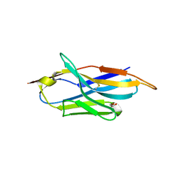 | | A llama-derived JBP1-targeting nanobody | | 分子名称: | GLYCEROL, Nanobody | | 著者 | van Beusekom, B, Adamopoulos, A, Heidebrecht, T, Joosten, R.P, Perrakis, A. | | 登録日 | 2018-02-12 | | 公開日 | 2018-10-31 | | 最終更新日 | 2024-11-13 | | 実験手法 | X-RAY DIFFRACTION (1.64 Å) | | 主引用文献 | Characterization and structure determination of a llama-derived nanobody targeting the J-base binding protein 1.
Acta Crystallogr F Struct Biol Commun, 74, 2018
|
|
3G73
 | | Structure of the FOXM1 DNA binding | | 分子名称: | DNA (5'-D(P*AP*AP*AP*TP*TP*GP*TP*TP*TP*AP*TP*AP*AP*AP*CP*AP*GP*CP*CP*CP*G)-3'), DNA (5'-D(P*TP*TP*CP*GP*GP*GP*CP*TP*GP*TP*TP*TP*AP*TP*AP*AP*AP*CP*AP*AP*T)-3'), Forkhead box protein M1, ... | | 著者 | Littler, D.R, Perrakis, A, Hibbert, R.G, Medema, R.H. | | 登録日 | 2009-02-09 | | 公開日 | 2009-03-03 | | 最終更新日 | 2023-11-01 | | 実験手法 | X-RAY DIFFRACTION (2.21 Å) | | 主引用文献 | Structure of the FoxM1 DNA-recognition domain bound to a promoter sequence
Nucleic Acids Res., 2010
|
|
5IJQ
 | | Crystal structure of autotaxin (ENPP2) re-refined | | 分子名称: | 7alpha-hydroxycholesterol, CALCIUM ION, Ectonucleotide pyrophosphatase/phosphodiesterase family member 2, ... | | 著者 | Hausmann, J, Joosten, R.P, Perrakis, A. | | 登録日 | 2016-03-02 | | 公開日 | 2016-06-15 | | 最終更新日 | 2024-11-13 | | 実験手法 | X-RAY DIFFRACTION (2.05 Å) | | 主引用文献 | Structural snapshots of the catalytic cycle of the phosphodiesterase Autotaxin.
J.Struct.Biol., 195, 2016
|
|
5IJS
 | | Crystal structure of autotaxin with orthovanadate bound as a trigonal bipyramidal intermediate analog | | 分子名称: | 2-acetamido-2-deoxy-beta-D-glucopyranose-(1-4)-2-acetamido-2-deoxy-beta-D-glucopyranose, 7alpha-hydroxycholesterol, CALCIUM ION, ... | | 著者 | Hausmann, J, Joosten, R.P, Perrakis, A. | | 登録日 | 2016-03-02 | | 公開日 | 2016-06-15 | | 最終更新日 | 2024-01-10 | | 実験手法 | X-RAY DIFFRACTION (2.2 Å) | | 主引用文献 | Structural snapshots of the catalytic cycle of the phosphodiesterase Autotaxin.
J.Struct.Biol., 195, 2016
|
|
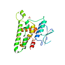
9QYF
 | | PARP9 Macro Domain 2 in complex with ADPr | | 分子名称: | ADENOSINE-5-DIPHOSPHORIBOSE, Protein mono-ADP-ribosyltransferase PARP9, [(2R,3S,4R,5R)-5-(6-AMINOPURIN-9-YL)-3,4-DIHYDROXY-OXOLAN-2-YL]METHYL[HYDROXY-[[(2R,3S,4R,5S)-3,4,5-TRIHYDROXYOXOLAN-2-YL]METHOXY]PHOSPHORYL] HYDROGEN PHOSPHATE | | 著者 | Fourkiotis, K.N, Chikunova, A, Tsika, C.A, Kravvariti, P.K, Tsatsouli, A.S, Perrakis, A, Spyroulias, A.G. | | 登録日 | 2025-04-17 | | 公開日 | 2025-05-28 | | 実験手法 | X-RAY DIFFRACTION (1.3 Å) | | 主引用文献 | Comparative analysis of hPARP9 macro domains
To Be Published
|
|
9QYH
 | | PARP9 Macro Domain 2 in complex with NAD+ | | 分子名称: | NICOTINAMIDE-ADENINE-DINUCLEOTIDE, Protein mono-ADP-ribosyltransferase PARP9 | | 著者 | Fourkiotis, K.N, Chikunova, A, Tsika, C.A, Kravvariti, P.K, Tsatsouli, A.S, Perrakis, A, Spyroulias, A.G. | | 登録日 | 2025-04-17 | | 公開日 | 2025-05-28 | | 実験手法 | X-RAY DIFFRACTION (2.5 Å) | | 主引用文献 | Comparative analysis of hPARP9 macro domains
To Be Published
|
|
9QYE
 | | PARP9 Macro domain 1 in complex with ADPr | | 分子名称: | Protein mono-ADP-ribosyltransferase PARP9, [(2R,3S,4R,5R)-5-(6-AMINOPURIN-9-YL)-3,4-DIHYDROXY-OXOLAN-2-YL]METHYL[HYDROXY-[[(2R,3S,4R,5S)-3,4,5-TRIHYDROXYOXOLAN-2-YL]METHOXY]PHOSPHORYL] HYDROGEN PHOSPHATE | | 著者 | Fourkiotis, K.N, Chikunova, A, Tsika, C.A, Tsatsouli, A.S, Perrakis, A, Spyroulias, A.G. | | 登録日 | 2025-04-17 | | 公開日 | 2025-05-28 | | 実験手法 | X-RAY DIFFRACTION (1.44 Å) | | 主引用文献 | Comparative analysis of hPARP9 macro domains
To Be Published
|
|
9QYG
 | | PARP9 Macro Domain 2 Free form | | 分子名称: | HEXAETHYLENE GLYCOL, Protein mono-ADP-ribosyltransferase PARP9 | | 著者 | Fourkiotis, K.N, Chikunova, A, Tsika, C.A, Kravvariti, P.K, Tsatsouli, A.S, Perrakis, A, Spyroulias, A.G. | | 登録日 | 2025-04-17 | | 公開日 | 2025-05-28 | | 実験手法 | X-RAY DIFFRACTION (2.7 Å) | | 主引用文献 | Comparative analysis of hPARP9 macro domains
To Be Published
|
|
9QYD
 | | PARP9 Macro domain 1, Free form | | 分子名称: | GLYCEROL, MALONATE ION, Protein mono-ADP-ribosyltransferase PARP9 | | 著者 | Fourkiotis, N.K, Chikunova, A, Tsika, C.A, Tsatsouli, A.S, Perrakis, A, Spyroulias, G.A. | | 登録日 | 2025-04-17 | | 公開日 | 2025-05-28 | | 実験手法 | X-RAY DIFFRACTION (1.91 Å) | | 主引用文献 | Comparative analysis of hPARP9 macro domains
To Be Published
|
|


