3BJH
 
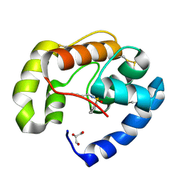 | | Soft-SAD crystal structure of a pheromone binding protein from the honeybee Apis mellifera L. | | 分子名称: | GLYCEROL, N-BUTYL-BENZENESULFONAMIDE, Pheromone-binding protein ASP1 | | 著者 | Lartigue, A, Gruez, A, Briand, L, Blon, F, Bezirard, V, Walsh, M, Pernollet, J.C, Tegoni, M, Cambillau, C. | | 登録日 | 2007-12-04 | | 公開日 | 2007-12-18 | | 最終更新日 | 2024-10-30 | | 実験手法 | X-RAY DIFFRACTION (1.6 Å) | | 主引用文献 | Sulfur single-wavelength anomalous diffraction crystal structure of a pheromone-binding protein from the honeybee Apis mellifera L.
J.Biol.Chem., 279, 2004
|
|
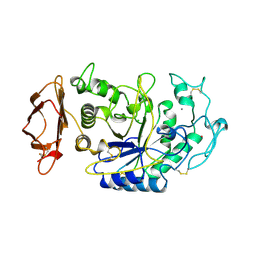
1JFH
 
 | |
1VAH
 
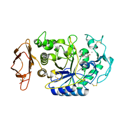 | | Crystal structure of the pig pancreatic-amylase complexed with r-nitrophenyl-a-D-maltoside | | 分子名称: | Alpha-amylase, pancreatic, CALCIUM ION, ... | | 著者 | Zhuo, H, Payan, F, Qian, M. | | 登録日 | 2004-02-17 | | 公開日 | 2005-04-26 | | 最終更新日 | 2023-12-27 | | 実験手法 | X-RAY DIFFRACTION (2.4 Å) | | 主引用文献 | Crystal structure of the pig pancreatic alpha-amylase complexed with rho-nitrophenyl-alpha-D-maltoside-flexibility in the active site
Protein J., 23, 2004
|
|
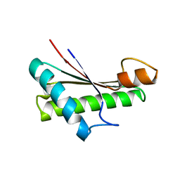
5M38
 
 | |
1N1T
 
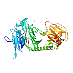 | | Trypanosoma rangeli sialidase in complex with DANA at 1.6 A | | 分子名称: | 2-DEOXY-2,3-DEHYDRO-N-ACETYL-NEURAMINIC ACID, SULFATE ION, Sialidase | | 著者 | Amaya, M.F, Buschiazzo, A, Nguyen, T, Alzari, P.M. | | 登録日 | 2002-10-20 | | 公開日 | 2003-01-07 | | 最終更新日 | 2024-11-06 | | 実験手法 | X-RAY DIFFRACTION (1.6 Å) | | 主引用文献 | The high resolution structures of free and
inhibitor-bound Trypanosoma rangeli
sialidase and its comparison with T. cruzi
trans-sialidase
J.Mol.Biol., 325, 2003
|
|
1N1S
 
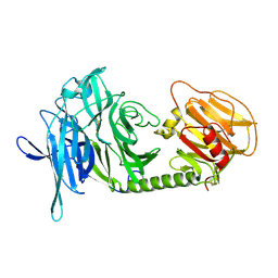 | | Trypanosoma rangeli sialidase | | 分子名称: | SULFATE ION, Sialidase | | 著者 | Amaya, M.F, Buschiazzo, A, Nguyen, T, Alzari, P.M. | | 登録日 | 2002-10-20 | | 公開日 | 2003-01-07 | | 最終更新日 | 2011-07-13 | | 実験手法 | X-RAY DIFFRACTION (1.64 Å) | | 主引用文献 | The high resolution structures of free and
inhibitor-bound Trypanosoma rangeli
sialidase and its comparison with T. cruzi
trans-sialidase
J.Mol.Biol., 325, 2003
|
|
1N1V
 
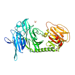 | | Trypanosoma rangeli sialidase in complex with DANA | | 分子名称: | 2-DEOXY-2,3-DEHYDRO-N-ACETYL-NEURAMINIC ACID, SULFATE ION, Sialidase | | 著者 | Amaya, M.F, Buschiazzo, A, Nguyen, T, Alzari, P.M. | | 登録日 | 2002-10-21 | | 公開日 | 2003-01-07 | | 最終更新日 | 2020-07-29 | | 実験手法 | X-RAY DIFFRACTION (2.1 Å) | | 主引用文献 | The high resolution structures of free and
inhibitor-bound Trypanosoma rangeli
sialidase and its comparison with T.
cruzi trans-sialidase
J.Mol.Biol., 325, 2003
|
|
1N1Y
 
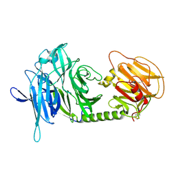 | | Trypanosoma rangeli sialidase in complex with sialic acid | | 分子名称: | N-acetyl-alpha-neuraminic acid, Sialidase | | 著者 | Amaya, M.F, Buschiazzo, A, Nguyen, T, Alzari, P.M. | | 登録日 | 2002-10-21 | | 公開日 | 2003-01-07 | | 最終更新日 | 2024-10-30 | | 実験手法 | X-RAY DIFFRACTION (2.8 Å) | | 主引用文献 | The high resolution structures of free and inhibitor-bound
Trypanosoma rangeli sialidase and its comparison with T.
cruzi trans-sialidase
J.Mol.Biol., 325, 2003
|
|
