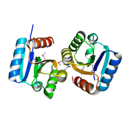
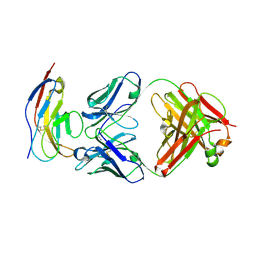
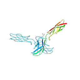
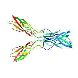
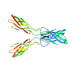
4ZPN
 
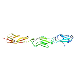 | | Crystal Structure of Protocadherin Gamma C5 EC1-3 with extended N-terminus | | 分子名称: | 2-acetamido-2-deoxy-beta-D-glucopyranose-(1-4)-[alpha-L-fucopyranose-(1-6)]2-acetamido-2-deoxy-beta-D-glucopyranose, CALCIUM ION, MCG133388, ... | | 著者 | Goodman, K.M, Wolcott, H.N, Bahna, F, Shapiro, L. | | 登録日 | 2015-05-08 | | 公開日 | 2015-10-28 | | 最終更新日 | 2023-09-27 | | 実験手法 | X-RAY DIFFRACTION (3.3 Å) | | 主引用文献 | Molecular Logic of Neuronal Self-Recognition through Protocadherin Domain Interactions.
Cell, 163, 2015
|
|
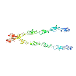
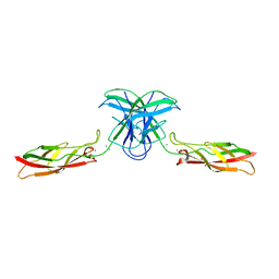
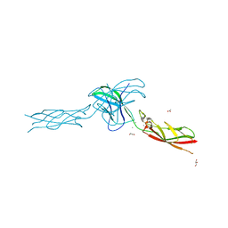
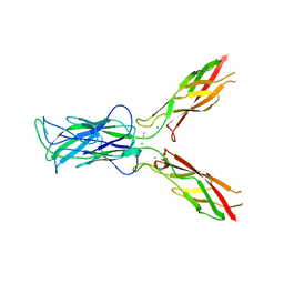
4ZPP
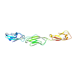 | | Crystal Structure of Protocadherin Gamma C5 EC1-3 | | 分子名称: | 2-acetamido-2-deoxy-beta-D-glucopyranose-(1-4)-2-acetamido-2-deoxy-beta-D-glucopyranose, CALCIUM ION, MCG133388, ... | | 著者 | Wolcott, H.N, Goodman, K.M, Bahna, F, Mannepalli, S, Shapiro, L. | | 登録日 | 2015-05-08 | | 公開日 | 2015-10-28 | | 最終更新日 | 2023-09-27 | | 実験手法 | X-RAY DIFFRACTION (3.002 Å) | | 主引用文献 | Molecular Logic of Neuronal Self-Recognition through Protocadherin Domain Interactions.
Cell, 163, 2015
|
|
8TNL
 | |
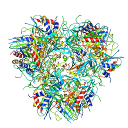
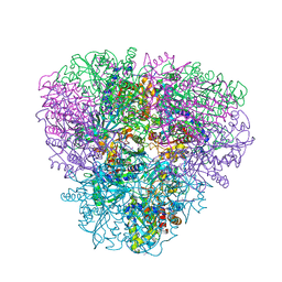
8TOA
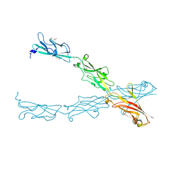
 | | CryoEM structure of H7 hemagglutinin from A/Shanghai2/2013 H7N9 in complex with a human neutralizing antibody H7.HK2 | | 分子名称: | 2-acetamido-2-deoxy-beta-D-glucopyranose, H7.HK2 Neutralizing Antibody Heavy Chain, H7.HK2 Neutralizing Antibody Light Chain, ... | | 著者 | Morano, N.C, Becker, J.E, Wu, X, Shapiro, L. | | 登録日 | 2023-08-03 | | 公開日 | 2024-05-15 | | 実験手法 | ELECTRON MICROSCOPY (3.69 Å) | | 主引用文献 | CryoEM structure of H7 hemagglutinin from A/Shanghai2/2013 H7N9 in complex with a human neutralizing antibody H7.HK1
To Be Published
|
|
4DN6
 | |
2IJR
 | | Crystal structure of a protein api92 from Yersinia pseudotuberculosis, Pfam DUF1281 | | 分子名称: | Hypothetical protein api92 | | 著者 | Jin, X, Min, T, Bonanno, J.B, Sauder, J.M, Wasserman, S, Smith, D, Burley, S.K, Shapiro, L, New York SGX Research Center for Structural Genomics (NYSGXRC) | | 登録日 | 2006-09-30 | | 公開日 | 2006-10-31 | | 最終更新日 | 2021-02-03 | | 実験手法 | X-RAY DIFFRACTION (2.7 Å) | | 主引用文献 | Crystal structure of a hypothetical protein from Yersinia
pseudotuberculosis
To be Published
|
|
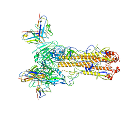
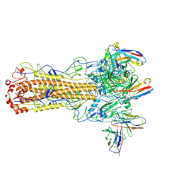
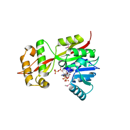
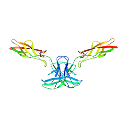
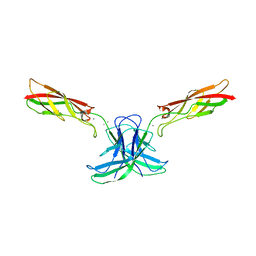
6E6B
 | | Crystal structure of the Protocadherin GammaB4 extracellular domain | | 分子名称: | 2-acetamido-2-deoxy-beta-D-glucopyranose, 2-acetamido-2-deoxy-beta-D-glucopyranose-(1-4)-2-acetamido-2-deoxy-beta-D-glucopyranose, 2-acetamido-2-deoxy-beta-D-glucopyranose-(1-4)-[alpha-L-fucopyranose-(1-6)]2-acetamido-2-deoxy-beta-D-glucopyranose, ... | | 著者 | Goodman, K.M, Mannepalli, S, Bahna, F, Honig, B, Shapiro, L. | | 登録日 | 2018-07-24 | | 公開日 | 2019-04-10 | | 最終更新日 | 2023-10-11 | | 実験手法 | X-RAY DIFFRACTION (4.52 Å) | | 主引用文献 | Visualization of clustered protocadherin neuronal self-recognition complexes.
Nature, 569, 2019
|
|
2IJZ
 | |
2GLU
 | | The crystal structure of YcgJ protein from Bacillus subitilis | | 分子名称: | S-ADENOSYLMETHIONINE, SULFATE ION, ycgJ | | 著者 | Burke, T, Gorman, J, Shapiro, L, Burley, S.K, New York SGX Research Center for Structural Genomics (NYSGXRC) | | 登録日 | 2006-04-05 | | 公開日 | 2006-04-18 | | 最終更新日 | 2023-11-15 | | 実験手法 | X-RAY DIFFRACTION (2.91 Å) | | 主引用文献 | The crystal structure of YcgJ protein from Bacillus subitilis
To be Published
|
|
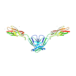
6BIT
 | | SIRPalpha antibody complex | | 分子名称: | KWAR23 Fab heavy chain, KWAR23 Fab light chain, Tyrosine-protein phosphatase non-receptor type substrate 1 | | 著者 | Ring, N.G, Herndler-Brandstetter, D, Weiskopf, K, Shan, L, Volkmer, J.P, George, B.M, Lietzenmayer, M, McKenna, K.M, Naik, T.J, McCarty, A, Zheng, Y, Ring, A.M, Flavell, R.A, Weissman, I.L. | | 登録日 | 2017-11-03 | | 公開日 | 2017-12-06 | | 最終更新日 | 2019-11-20 | | 実験手法 | X-RAY DIFFRACTION (2.191 Å) | | 主引用文献 | Anti-SIRP alpha antibody immunotherapy enhances neutrophil and macrophage antitumor activity.
Proc. Natl. Acad. Sci. U.S.A., 114, 2017
|
|
6CG7
 | |
6CGS
 | | mouse cadherin-7 EC1-2 adhesive fragment | | 分子名称: | CALCIUM ION, Cadherin-7, GLYCEROL | | 著者 | Brasch, J, Harrison, O.J, Kaczynska, A, Shapiro, L. | | 登録日 | 2018-02-20 | | 公開日 | 2018-05-09 | | 最終更新日 | 2023-10-04 | | 実験手法 | X-RAY DIFFRACTION (1.72 Å) | | 主引用文献 | Homophilic and Heterophilic Interactions of Type II Cadherins Identify Specificity Groups Underlying Cell-Adhesive Behavior.
Cell Rep, 23, 2018
|
|
2I5G
 | | Crystal strcuture of amidohydrolase from Pseudomonas aeruginosa | | 分子名称: | amidohydrolase | | 著者 | Min, T, Sauder, J.M, Wasserman, S.R, Smith, D, Burley, S.K, Shapiro, L, New York SGX Research Center for Structural Genomics (NYSGXRC) | | 登録日 | 2006-08-24 | | 公開日 | 2006-09-05 | | 最終更新日 | 2021-10-20 | | 実験手法 | X-RAY DIFFRACTION (2.6 Å) | | 主引用文献 | Crystal structure of amidohydrolase from Pseudomonas aeruginosa
To be Published
|
|
6CV7
 | |
6CG6
 | | mouse cadherin-10 EC1-2 adhesive fragment | | 分子名称: | 1,2-ETHANEDIOL, CALCIUM ION, Cadherin-10, ... | | 著者 | Brasch, J, Harrison, O.J, Shapiro, L. | | 登録日 | 2018-02-19 | | 公開日 | 2018-05-09 | | 最終更新日 | 2023-10-04 | | 実験手法 | X-RAY DIFFRACTION (2.707 Å) | | 主引用文献 | Homophilic and Heterophilic Interactions of Type II Cadherins Identify Specificity Groups Underlying Cell-Adhesive Behavior.
Cell Rep, 23, 2018
|
|
6CGU
 | |
6CGB
 | | chimera of mouse cadherin-11 EC1 and mouse cadherin-6 EC2 | | 分子名称: | ACETATE ION, CALCIUM ION, Cadherin-11, ... | | 著者 | Brasch, J, Harrison, O.J, Shapiro, L, Kaeser, B. | | 登録日 | 2018-02-19 | | 公開日 | 2018-05-09 | | 最終更新日 | 2023-10-04 | | 実験手法 | X-RAY DIFFRACTION (2.994 Å) | | 主引用文献 | Homophilic and Heterophilic Interactions of Type II Cadherins Identify Specificity Groups Underlying Cell-Adhesive Behavior.
Cell Rep, 23, 2018
|
|
2GLJ
 | |
2GLF
 | |
2KHS
 | | Solution structure of SNase121:SNase(111-143) complex | | 分子名称: | Nuclease, Thermonuclease | | 著者 | Geng, Y, Feng, Y, Xie, T, Shan, L, Wang, J. | | 登録日 | 2009-04-10 | | 公開日 | 2009-10-20 | | 最終更新日 | 2024-05-15 | | 実験手法 | SOLUTION NMR | | 主引用文献 | The native-like interactions between SNase121 and SNase(111-143) fragments induce the recovery of their native-like structures and the ability to degrade DNA.
Biochemistry, 48, 2009
|
|
4NQQ
 | |
3LNE
 | |
3LNH
 | |
3LND
 | |
3LNG
 | |


