3A6V
 
 | |
3A6T
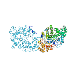
 | | Crystal structure of MutT-8-OXO-DGMP complex | | 分子名称: | 8-OXO-2'-DEOXY-GUANOSINE-5'-MONOPHOSPHATE, Mutator mutT protein, SODIUM ION, ... | | 著者 | Nakamura, T, Yamagata, Y. | | 登録日 | 2009-09-09 | | 公開日 | 2009-10-27 | | 最終更新日 | 2023-11-01 | | 実験手法 | X-RAY DIFFRACTION (1.96 Å) | | 主引用文献 | Structural and dynamic features of the MutT protein in the recognition of nucleotides with the mutagenic 8-oxoguanine base
J.Biol.Chem., 285, 2010
|
|
3A6U
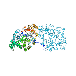
 | | Crystal structure of MutT-8-OXO-dGMP-MN(II) complex | | 分子名称: | 8-OXO-2'-DEOXY-GUANOSINE-5'-MONOPHOSPHATE, MANGANESE (II) ION, Mutator mutT protein, ... | | 著者 | Nakamura, T, Yamagata, Y. | | 登録日 | 2009-09-09 | | 公開日 | 2009-10-27 | | 最終更新日 | 2023-11-01 | | 実験手法 | X-RAY DIFFRACTION (2.56 Å) | | 主引用文献 | Structural and dynamic features of the MutT protein in the recognition of nucleotides with the mutagenic 8-oxoguanine base
J.Biol.Chem., 285, 2010
|
|
3A6S
 | | Crystal structure of the MutT protein | | 分子名称: | L(+)-TARTARIC ACID, Mutator mutT protein, SODIUM ION | | 著者 | Nakamura, T, Yamagata, Y. | | 登録日 | 2009-09-09 | | 公開日 | 2009-10-27 | | 最終更新日 | 2024-03-13 | | 実験手法 | X-RAY DIFFRACTION (1.8 Å) | | 主引用文献 | Structural and dynamic features of the MutT protein in the recognition of nucleotides with the mutagenic 8-oxoguanine base
J.Biol.Chem., 285, 2010
|
|
3A1M
 | | A fusion protein of a beta helix region of gene product 5 and the foldon region of bacteriophage T4 | | 分子名称: | POTASSIUM ION, chimera of thrombin cleavage site, Tail-associated lysozyme, ... | | 著者 | Yokoi, N, Suzuki, A, Hikage, T, Koshiyama, T, Terauchi, M, Yutani, K, Kanamaru, S, Arisaka, F, Yamane, T, Watanabe, Y, Ueno, T. | | 登録日 | 2009-04-11 | | 公開日 | 2010-04-21 | | 最終更新日 | 2023-11-01 | | 実験手法 | X-RAY DIFFRACTION (2 Å) | | 主引用文献 | Construction of Robust Bio-nanotubes using the Controlled Self-Assembly of Component Proteins of Bacteriophage T4
Small, 6, 2010
|
|
8EE2
 | |
8E1C
 | |
8E1B
 | |
8E2N
 | |
8EVD
 | |
8F6U
 | |
8F6V
 | |
8GJR
 | |
7COV
 | | Potato D-enzyme, native (substrate free) | | 分子名称: | 4-alpha-glucanotransferase, chloroplastic/amyloplastic, CALCIUM ION, ... | | 著者 | Unno, H, Imamura, K. | | 登録日 | 2020-08-05 | | 公開日 | 2020-08-26 | | 最終更新日 | 2024-03-27 | | 実験手法 | X-RAY DIFFRACTION (2 Å) | | 主引用文献 | Structural analysis and reaction mechanism of the disproportionating enzyme (D-enzyme) from potato.
Protein Sci., 29, 2020
|
|
6LX2
 | | Potato D-enzyme complexed with CA26 | | 分子名称: | 4-alpha-glucanotransferase, chloroplastic/amyloplastic, 4-deoxy-alpha-D-glucopyranose-(1-4)-4-deoxy-alpha-D-glucopyranose-(1-4)-4-deoxy-alpha-D-glucopyranose-(1-4)-4-deoxy-alpha-D-glucopyranose-(1-4)-4-deoxy-alpha-D-glucopyranose-(1-4)-4-deoxy-alpha-D-glucopyranose-(1-4)-4-deoxy-alpha-D-glucopyranose-(1-4)-4-deoxy-alpha-D-glucopyranose-(1-4)-4-deoxy-alpha-D-glucopyranose-(1-4)-4-deoxy-alpha-D-glucopyranose-(1-4)-4-deoxy-alpha-D-glucopyranose, ... | | 著者 | Unno, H, Imamura, K. | | 登録日 | 2020-02-10 | | 公開日 | 2020-08-26 | | 最終更新日 | 2023-11-29 | | 実験手法 | X-RAY DIFFRACTION (2.05 Å) | | 主引用文献 | Structural analysis and reaction mechanism of the disproportionating enzyme (D-enzyme) from potato.
Protein Sci., 29, 2020
|
|
6LX1
 | | Potato D-enzyme complexed with Acarbose | | 分子名称: | 4,6-dideoxy-4-{[(1S,4R,5S,6S)-4,5,6-trihydroxy-3-(hydroxymethyl)cyclohex-2-en-1-yl]amino}-alpha-D-glucopyranose-(1-4)-1,5-anhydro-D-glucitol, 4-alpha-glucanotransferase, chloroplastic/amyloplastic, ... | | 著者 | Unno, H, Imamura, K. | | 登録日 | 2020-02-10 | | 公開日 | 2020-08-26 | | 最終更新日 | 2023-11-29 | | 実験手法 | X-RAY DIFFRACTION (2.03 Å) | | 主引用文献 | Structural analysis and reaction mechanism of the disproportionating enzyme (D-enzyme) from potato.
Protein Sci., 29, 2020
|
|
3VKN
 | |
1I1Z
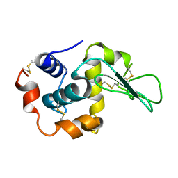 | | MUTANT HUMAN LYSOZYME (Q86D) | | 分子名称: | LYSOZYME C | | 著者 | Kuroki, R. | | 登録日 | 2001-02-05 | | 公開日 | 2001-02-28 | | 最終更新日 | 2024-04-03 | | 実験手法 | X-RAY DIFFRACTION (1.8 Å) | | 主引用文献 | Structural and thermodynamic responses of mutations at a Ca2+ binding site engineered into human lysozyme.
J.Biol.Chem., 273, 1998
|
|
1I20
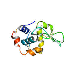
 | | MUTANT HUMAN LYSOZYME (A92D) | | 分子名称: | LYSOZYME C | | 著者 | Kuroki, R. | | 登録日 | 2001-02-05 | | 公開日 | 2001-02-28 | | 最終更新日 | 2023-10-25 | | 実験手法 | X-RAY DIFFRACTION (1.9 Å) | | 主引用文献 | Structural and thermodynamic responses of mutations at a Ca2+ binding site engineered into human lysozyme.
J.Biol.Chem., 273, 1998
|
|
1I22
 | | MUTANT HUMAN LYSOZYME (A83K/Q86D/A92D) | | 分子名称: | CALCIUM ION, LYSOZYME C | | 著者 | Kuroki, R. | | 登録日 | 2001-02-05 | | 公開日 | 2001-02-28 | | 最終更新日 | 2023-10-25 | | 実験手法 | X-RAY DIFFRACTION (1.8 Å) | | 主引用文献 | Structural and thermodynamic responses of mutations at a Ca2+ binding site engineered into human lysozyme.
J.Biol.Chem., 273, 1998
|
|
3VKL
 | |
3VKO
 | | Galectin-8 N-terminal domain in complex with sialyllactosamine | | 分子名称: | CHLORIDE ION, Galectin-8, N-acetyl-alpha-neuraminic acid-(2-3)-beta-D-galactopyranose-(1-4)-2-acetamido-2-deoxy-beta-D-glucopyranose | | 著者 | Kamitori, S, Yoshida, H. | | 登録日 | 2011-11-18 | | 公開日 | 2012-09-12 | | 最終更新日 | 2023-11-08 | | 実験手法 | X-RAY DIFFRACTION (2.08 Å) | | 主引用文献 | X-ray structure of a protease-resistant mutant form of human galectin-8 with two carbohydrate recognition domains
Febs J., 279, 2012
|
|
2ZU0
 | |
3WNO
 | | D308A mutant of Bacillus circulans T-3040 cycloisomaltooligosaccharide glucanotransferase complexed with cycloisomaltooctaose | | 分子名称: | 1,2-ETHANEDIOL, 2-(N-MORPHOLINO)-ETHANESULFONIC ACID, CALCIUM ION, ... | | 著者 | Suzuki, N, Fujimoto, Z, Kim, Y.M, Momma, M, Kishine, N, Suzuki, R, Kobayashi, M, Kimura, A, Funane, K. | | 登録日 | 2013-12-10 | | 公開日 | 2014-02-05 | | 最終更新日 | 2023-11-08 | | 実験手法 | X-RAY DIFFRACTION (1.9 Å) | | 主引用文献 | Structural elucidation of the cyclization mechanism of alpha-1,6-glucan by Bacillus circulans T-3040 cycloisomaltooligosaccharide glucanotransferase.
J.Biol.Chem., 289, 2014
|
|
3WUS
 | | Crystal Structure of the Vif-Binding Domain of Human APOBEC3F | | 分子名称: | DNA dC->dU-editing enzyme APOBEC-3F, ZINC ION | | 著者 | Nakashima, M, Kawamura, T, Ode, H, Watanabe, N, Iwatani, Y. | | 登録日 | 2014-05-02 | | 公開日 | 2015-06-24 | | 最終更新日 | 2023-11-08 | | 実験手法 | X-RAY DIFFRACTION (2.54 Å) | | 主引用文献 | Structural Insights into HIV-1 Vif-APOBEC3F Interaction.
J.Virol., 90, 2015
|
|


