2GH2
 
 | |
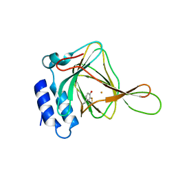
1QFY
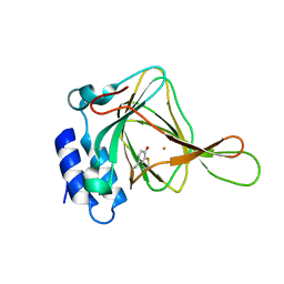
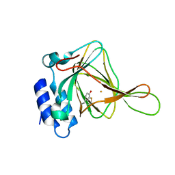
 | | PEA FNR Y308S MUTANT IN COMPLEX WITH NADP+ | | 分子名称: | FLAVIN-ADENINE DINUCLEOTIDE, NADP NICOTINAMIDE-ADENINE-DINUCLEOTIDE PHOSPHATE, PROTEIN (FERREDOXIN: NADP+ REDUCTASE), ... | | 著者 | Deng, Z, Aliverti, A, Zanetti, G, Arakaki, A.K, Ottado, J, Orellano, E.G, Calcaterra, N.B, Ceccarelli, E.A, Carrillo, N, Karplus, P.A. | | 登録日 | 1999-04-18 | | 公開日 | 1999-04-27 | | 最終更新日 | 2024-04-03 | | 実験手法 | X-RAY DIFFRACTION (1.8 Å) | | 主引用文献 | A productive NADP+ binding mode of ferredoxin-NADP + reductase revealed by protein engineering and crystallographic studies.
Nat.Struct.Biol., 6, 1999
|
|
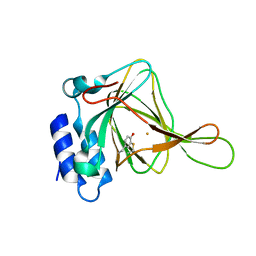
1QG0
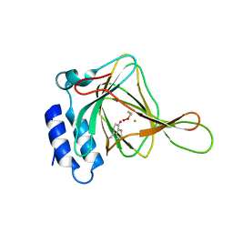
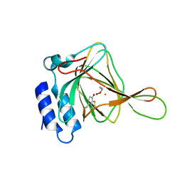
 | | WILD-TYPE PEA FNR | | 分子名称: | FLAVIN-ADENINE DINUCLEOTIDE, PROTEIN (FERREDOXIN:NADP+ REDUCTASE) | | 著者 | Deng, Z, Aliverti, A, Zanetti, G, Arakaki, A.K, Ottado, J, Orellano, E.G, Calcaterra, N.B, Ceccarelli, E.A, Carrillo, N, Karplus, P.A. | | 登録日 | 1999-04-18 | | 公開日 | 1999-04-27 | | 最終更新日 | 2023-08-16 | | 実験手法 | X-RAY DIFFRACTION (2.5 Å) | | 主引用文献 | A productive NADP+ binding mode of ferredoxin-NADP+ reductase revealed by protein engineering and crystallographic studies.
Nat.Struct.Biol., 6, 1999
|
|
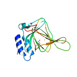
1QGA
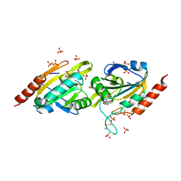
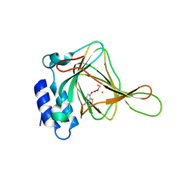
 | | PEA FNR Y308W MUTANT IN COMPLEX WITH NADP+ | | 分子名称: | FLAVIN-ADENINE DINUCLEOTIDE, NADP NICOTINAMIDE-ADENINE-DINUCLEOTIDE PHOSPHATE, PROTEIN (FERREDOXIN:NADP+ REDUCTASE), ... | | 著者 | Deng, Z, Aliverti, A, Zanetti, G, Arakaki, A.K, Ottado, J, Orellano, E.G, Calcaterra, N.B, Ceccarelli, E.A, Carrillo, N, Karplus, P.A. | | 登録日 | 1999-04-18 | | 公開日 | 1999-04-27 | | 最終更新日 | 2024-12-25 | | 実験手法 | X-RAY DIFFRACTION (2 Å) | | 主引用文献 | A productive NADP+ binding mode of ferredoxin-NADP+ reductase revealed by protein engineering and crystallographic studies.
Nat.Struct.Biol., 6, 1999
|
|
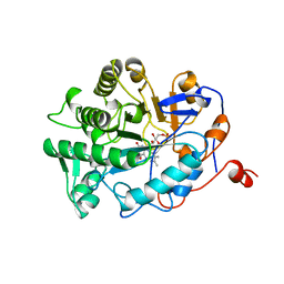
1SGH
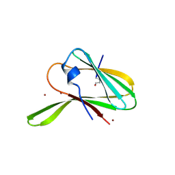
 | | Moesin FERM domain bound to EBP50 C-terminal peptide | | 分子名称: | Ezrin-radixin-moesin binding phosphoprotein 50, Moesin | | 著者 | Finnerty, C.M, Chambers, D, Ingraffea, J, Faber, H.R, Karplus, P.A, Bretscher, A. | | 登録日 | 2004-02-23 | | 公開日 | 2004-06-29 | | 最終更新日 | 2023-08-23 | | 実験手法 | X-RAY DIFFRACTION (3.5 Å) | | 主引用文献 | The EBP50-moesin interaction involves a binding site regulated by direct masking on the FERM domain
J.Cell.Sci., 117, 2004
|
|
1QFZ
 | | PEA FNR Y308S MUTANT IN COMPLEX WITH NADPH | | 分子名称: | FLAVIN-ADENINE DINUCLEOTIDE, NADPH DIHYDRO-NICOTINAMIDE-ADENINE-DINUCLEOTIDE PHOSPHATE, PROTEIN (FERREDOXIN:NADP+ REDUCTASE), ... | | 著者 | Deng, Z, Aliverti, A, Zanetti, G, Arakaki, A.K, Ottado, J, Orellano, E.G, Calcaterra, N.B, Ceccarelli, E.A, Carrillo, N, Karplus, P.A. | | 登録日 | 1999-04-18 | | 公開日 | 1999-04-27 | | 最終更新日 | 2024-04-03 | | 実験手法 | X-RAY DIFFRACTION (1.7 Å) | | 主引用文献 | A productive NADP+ binding mode of ferredoxin-NADP+ reductase revealed by protein engineering and crystallographic studies.
Nat.Struct.Biol., 6, 1999
|
|
1TF4
 | |
4IES
 | | Cys-persulfenate bound Cysteine Dioxygenase at pH 6.2 in the presence of Cys | | 分子名称: | Cysteine dioxygenase type 1, FE (III) ION, S-HYDROPEROXYCYSTEINE | | 著者 | Driggers, C.M, Cooley, R.B, Sankaran, B, Karplus, P.A. | | 登録日 | 2012-12-13 | | 公開日 | 2013-06-26 | | 最終更新日 | 2024-11-06 | | 実験手法 | X-RAY DIFFRACTION (1.4 Å) | | 主引用文献 | Cysteine Dioxygenase Structures from pH4 to 9: Consistent Cys-Persulfenate Formation at Intermediate pH and a Cys-Bound Enzyme at Higher pH.
J.Mol.Biol., 425, 2013
|
|
4IEW
 | |
4IEX
 | |
4IEQ
 | |
4IET
 | |
4IEY
 | | Cys-persulfenate bound Cysteine Dioxygenase at pH 7.0 in the presence of Cys, home-source structure | | 分子名称: | Cysteine dioxygenase type 1, FE (II) ION, S-HYDROPEROXYCYSTEINE | | 著者 | Driggers, C.M, Cooley, R.B, Karplus, P.A. | | 登録日 | 2012-12-13 | | 公開日 | 2013-06-26 | | 最終更新日 | 2024-11-27 | | 実験手法 | X-RAY DIFFRACTION (1.63 Å) | | 主引用文献 | Cysteine Dioxygenase Structures from pH4 to 9: Consistent Cys-Persulfenate Formation at Intermediate pH and a Cys-Bound Enzyme at Higher pH.
J.Mol.Biol., 425, 2013
|
|
4GQC
 | | Crystal Structure of Aeropyrum pernix Peroxiredoxin Q Enzyme in Fully-Folded and Locally-Unfolded Conformations | | 分子名称: | DITHIANE DIOL, GLYCEROL, SULFATE ION, ... | | 著者 | Perkins, A, Karplus, P.A, Gretes, M.C, Nelson, K.J, Poole, L.B. | | 登録日 | 2012-08-22 | | 公開日 | 2012-10-24 | | 最終更新日 | 2024-11-27 | | 実験手法 | X-RAY DIFFRACTION (2 Å) | | 主引用文献 | Mapping the Active Site Helix-to-Strand Conversion of CxxxxC Peroxiredoxin Q Enzymes.
Biochemistry, 51, 2012
|
|
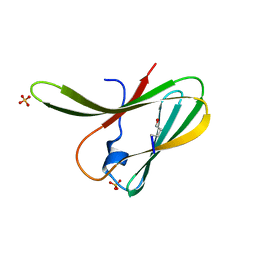
1ZLE
 | | Crystal structure of a RGD-containing host-selective toxin: Pyrenophora tritici-repentis Ptr ToxA | | 分子名称: | NICKEL (II) ION, Ptr necrosis toxin | | 著者 | Sarma, G.N, Manning, V.A, Ciuffetti, L.M, Karplus, P.A. | | 登録日 | 2005-05-06 | | 公開日 | 2005-08-16 | | 最終更新日 | 2024-10-30 | | 実験手法 | X-RAY DIFFRACTION (1.9 Å) | | 主引用文献 | Structure of Ptr ToxA: An RGD-Containing Host-Selective Toxin from Pyrenophora tritici-repentis
Plant Cell, 17, 2005
|
|
1ZLD
 | | Crystal structure of a RGD-containing host-selective toxin: Pyrenophora tritici-repentis Ptr ToxA | | 分子名称: | Ptr necrosis toxin, SULFATE ION | | 著者 | Sarma, G.N, Manning, V.A, Ciuffetti, L.M, Karplus, P.A. | | 登録日 | 2005-05-06 | | 公開日 | 2005-08-16 | | 最終更新日 | 2024-11-06 | | 実験手法 | X-RAY DIFFRACTION (1.65 Å) | | 主引用文献 | Structure of Ptr ToxA: An RGD-Containing Host-Selective Toxin from Pyrenophora tritici-repentis
Plant Cell, 17, 2005
|
|
4IEP
 | |
4IEU
 | |
4IER
 | |
4IEZ
 | |
4IEO
 | |
4IEV
 | |
1BWK
 | | OLD YELLOW ENZYME (OYE1) MUTANT H191N | | 分子名称: | FLAVIN MONONUCLEOTIDE, PROTEIN (NADPH DEHYDROGENASE 1) | | 著者 | Brown, B.J, Deng, Z, Karplus, P.A, Massey, V. | | 登録日 | 1998-09-24 | | 公開日 | 1998-09-30 | | 最終更新日 | 2023-08-09 | | 実験手法 | X-RAY DIFFRACTION (2.3 Å) | | 主引用文献 | On the active site of Old Yellow Enzyme. Role of histidine 191 and asparagine 194.
J.Biol.Chem., 273, 1998
|
|
1BWL
 | | OLD YELLOW ENZYME (OYE1) DOUBLE MUTANT H191N:N194H | | 分子名称: | FLAVIN MONONUCLEOTIDE, PROTEIN (NADPH DEHYDROGENASE 1) | | 著者 | Brown, B.J, Deng, Z, Karplus, P.A, Massey, V. | | 登録日 | 1998-09-24 | | 公開日 | 1998-09-30 | | 最終更新日 | 2023-08-09 | | 実験手法 | X-RAY DIFFRACTION (2.7 Å) | | 主引用文献 | On the active site of Old Yellow Enzyme. Role of histidine 191 and asparagine 194.
J.Biol.Chem., 273, 1998
|
|
1ZYN
 | | Oxidized structure of the N-terminal domain of Salmonella typhimurium AhpF | | 分子名称: | Alkyl hydroperoxide reductase subunit F | | 著者 | Roberts, B.R, Wood, Z.A, Jonsson, T.J, Poole, L.B, Karplus, P.A. | | 登録日 | 2005-06-10 | | 公開日 | 2005-06-21 | | 最終更新日 | 2024-10-16 | | 実験手法 | X-RAY DIFFRACTION (2.3 Å) | | 主引用文献 | Oxidized and synchrotron cleaved structures of the disulfide redox center in the N-terminal domain of Salmonella typhimurium AhpF
Protein Sci., 14, 2005
|
|


