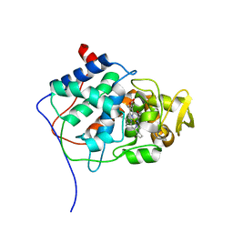
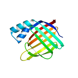
Loading
PDBj
メニューPDBj@FacebookPDBj@TwitterPDBj@YouTubewwPDB FoundationwwPDB
RCSB PDBPDBeBMRBAdv. SearchSearch help
Search by PDB author
1BJ9
DownloadVisualize
BU of 1bj9 by Molmil
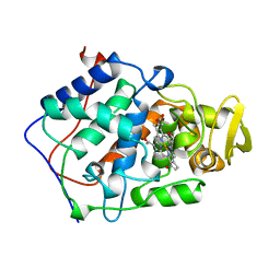
EFFECT OF UNNATURAL HEME SUBSTITUTION ON KINETICS OF ELECTRON TRANSFER IN CYTOCHROME C PEROXIDASE
分子名称: CYTOCHROME C PEROXIDASE, [7,12-DEACETYL-3,8,13,17-TETRAMETHYL-21H,23H-PORPHINE-2,18-DIPROPANOATO(2-)-N21,N22,N23,N24]-IRON
著者Miller, M.A, Kraut, J.
登録日1998-07-03
公開日1999-01-13
最終更新日2024-02-07
実験手法X-RAY DIFFRACTION (2.2 Å)
主引用文献Effect of Unnatural Heme Substitution on Kinetics of Electron Transfer in Cytochrome C Peroxidase
To be Published
4YGG
DownloadVisualize
BU of 4ygg by Molmil
CRYSTAL STRUCTURE OF R111K:Y134F:T54V:R132Q:P39Y:R59Y MUTANT OF HUMAN CELLULAR RETINOIC ACID BINDING PROTEIN II WITH RETINAL AT 1.9 ANGSTROM RESOLUTION - VISIBLE LIGHT IRRADIATED CRYSTAL - 2ND CYCLE
分子名称: Cellular retinoic acid-binding protein 2, RETINAL
著者Nosrati, M, Geiger, J.H.
登録日2015-02-26
公開日2016-03-02
最終更新日2023-09-27
実験手法X-RAY DIFFRACTION (1.9 Å)
主引用文献A Photoisomerizing Rhodopsin Mimic Observed at Atomic Resolution.
J.Am.Chem.Soc., 138, 2016
4YBU
DownloadVisualize
BU of 4ybu by Molmil
Crystal structure of the R111K:Y134F:T54V:R132Q:P39Q:R59Y mutant of human Cellular Retinoic Acid Binding ProteinII in complex with Retinal after 24 h incubation and 1 hour UV irradiation at 1.92 angstrom - 1st cycle
分子名称: Cellular retinoic acid-binding protein 2, RETINAL
著者Nosrati, M, Geiger, J.H.
登録日2015-02-19
公開日2016-02-24
最終更新日2023-09-27
実験手法X-RAY DIFFRACTION (1.924 Å)
主引用文献A Photoisomerizing Rhodopsin Mimic Observed at Atomic Resolution.
J.Am.Chem.Soc., 138, 2016
4YCH
DownloadVisualize
BU of 4ych by Molmil
Crystal Structure of R111K:Y134F:T54V:R132Q:P39Q:R59Y mutant of human Cellular Retinoic Acid Binding ProteinII with Retinal at 1.96 Angstrom - UV irradiated Crystal for 1 hour - 2nd cycle
分子名称: Cellular retinoic acid-binding protein 2, RETINAL
著者Nosrati, M, Geiger, J.H.
登録日2015-02-20
公開日2016-02-24
最終更新日2023-09-27
実験手法X-RAY DIFFRACTION (1.96 Å)
主引用文献A Photoisomerizing Rhodopsin Mimic Observed at Atomic Resolution.
J.Am.Chem.Soc., 138, 2016
1CYF
DownloadVisualize
BU of 1cyf by Molmil
IDENTIFYING THE PHYSIOLOGICAL ELECTRON TRANSFER SITE OF CYTOCHROME C PEROXIDASE BY STRUCTURE-BASED ENGINEERING
分子名称: CYTOCHROME C PEROXIDASE, PROTOPORPHYRIN IX CONTAINING FE
著者Miller, M.A, Han, G.W, Kraut, J.
登録日1995-07-03
公開日1995-12-07
最終更新日2024-02-07
実験手法X-RAY DIFFRACTION (2.35 Å)
主引用文献Identifying the physiological electron transfer site of cytochrome c peroxidase by structure-based engineering.
Biochemistry, 35, 1996
5F58
DownloadVisualize
BU of 5f58 by Molmil
Crystal structure of the Q108K:K40L:T51V:R58F mutant of human Cellular Retinol Binding Protein II in complex with All-trans-Retinal after 24 hours of incubation at 1.54 Angstrom Resolution
分子名称: ACETATE ION, RETINAL, Retinol-binding protein 2
著者Nosrati, M, Nossoni, Z, Geiger, J.H.
登録日2015-12-04
公開日2016-12-07
最終更新日2023-09-27
実験手法X-RAY DIFFRACTION (1.54 Å)
主引用文献Crystal structure of the Q108K:K40L:T51V:R58F mutant of human Cellular Retinol Binding Protein II in complex with All-trans-Retinal after 24 hours of incubation at 1.54 Angstrom Resolution
To Be Published
5F6B
DownloadVisualize
BU of 5f6b by Molmil
Crystal structure of the Q108K:K40L:T51V:R58Y:Y19W mutant of human Cellular Retinol Binding Protein II in complex with All-trans-Retinal at 1.3 Angstrom Resolution
分子名称: ACETATE ION, RETINAL, Retinol-binding protein 2
著者Nosrati, M, Nossoni, Z, Geiger, J.H.
登録日2015-12-05
公開日2016-12-07
最終更新日2023-09-27
実験手法X-RAY DIFFRACTION (1.31 Å)
主引用文献Crystal structure of the Q108K:K40L:T51V:R58Y:Y19W mutant of human Cellular Retinol Binding ProteinII in complex with All-trans-Retinal at 1.31 Angstrom Resolution
To Be Published
5FAZ
DownloadVisualize
BU of 5faz by Molmil
Crystal structure of the Q108K:K40L:T51V mutant of human Cellular Retinol Binding Protein II in complex with All-trans-Retinal after 24 hours of incubation at 1.4 Angstrom Resolution
分子名称: ACETATE ION, RETINAL, Retinol-binding protein 2
著者Nosrati, M, Nossoni, Z, Geiger, J.H.
登録日2015-12-13
公開日2016-12-14
最終更新日2023-09-27
実験手法X-RAY DIFFRACTION (1.4 Å)
主引用文献Crystal structure of the Q108K:K40L:T51V mutant of human Cellular Retinol Binding Protein II in complex with All-trans-Retinal after 24 hours of incubation at 1.54 Angstrom Resolution
To Be Published
5FEN
DownloadVisualize
BU of 5fen by Molmil
Crystal structure of the Q108K:K40L:T53C mutant of human Cellular Retinol Binding Protein II in complex with All-trans-Retinal after 24 hours of incubation at 1.55 Angstrom Resolution
分子名称: ACETATE ION, RETINAL, Retinol-binding protein 2
著者Nossoni, Z, Nosrati, M, Geiger, J.H.
登録日2015-12-17
公開日2016-12-21
最終更新日2023-09-27
実験手法X-RAY DIFFRACTION (1.55 Å)
主引用文献Crystal structure of the Q108K:K40L:T53C mutant of human Cellular Retinol Binding Protein II in complex with All-trans-Retinal after 24 hours of incubation at 1.55 Angstrom Resolution
To Be Published
5F7G
DownloadVisualize
BU of 5f7g by Molmil
Crystal structure of the Q108K:K40L:T51V:R58Y:Y19W:Q38L mutant of human Cellular Retinol Binding Protein II in complex with All-trans-Retinal at 1.48 Angstrom Resolution
分子名称: ACETATE ION, RETINAL, Retinol-binding protein 2
著者Nosrati, M, Nossoni, Z, Geiger, J.H.
登録日2015-12-08
公開日2016-12-14
最終更新日2023-09-27
実験手法X-RAY DIFFRACTION (1.48 Å)
主引用文献Crystal structure of the Q108K:K40L:T51V:R58F mutant of human Cellular Retinol Binding Protein II in complex with All-trans-Retinal after 24 hours of incubation at 1.54 Angstrom Resolution
To Be Published
5FFH
DownloadVisualize
BU of 5ffh by Molmil
Crystal structure of the Q108K:K40L:T51V:R58W:Y19W mutant of human Cellular Retinol Binding Protein II in complex with All-trans-Retinal at 1.68 Angstrom Resolution
分子名称: ACETATE ION, RETINAL, Retinol-binding protein 2
著者Nossoni, Z, Nosrati, M, Geiger, J.H.
登録日2015-12-18
公開日2016-12-21
最終更新日2023-09-27
実験手法X-RAY DIFFRACTION (1.68 Å)
主引用文献Crystal structure of the Q108K:K40L:T51V:R58W:Y19W mutant of human Cellular Retinol Binding ProteinII in complex with All-trans-Retinal at 1.68 Angstrom Resolution
To Be Published
3EDI
DownloadVisualize
BU of 3edi by Molmil
Crystal structure of tolloid-like protease 1 (TLL-1) protease domain
分子名称: ACETYL GROUP, DIMETHYL SULFOXIDE, SULFATE ION, ...
著者Mac Sweeney, A.
登録日2008-09-03
公開日2008-09-16
最終更新日2023-11-01
実験手法X-RAY DIFFRACTION (1.4 Å)
主引用文献Structural basis for the substrate specificity of bone morphogenetic protein 1/tolloid-like metalloproteases
To be Published
3EDH
DownloadVisualize
BU of 3edh by Molmil
Crystal structure of bone morphogenetic protein 1 protease domain in complex with partially bound DMSO
分子名称: ACETYL GROUP, Bone morphogenetic protein 1, DIMETHYL SULFOXIDE, ...
著者Mac Sweeney, A.
登録日2008-09-03
公開日2008-09-16
最終更新日2023-11-01
実験手法X-RAY DIFFRACTION (1.25 Å)
主引用文献Structural basis for the substrate specificity of bone morphogenetic protein 1/tolloid-like metalloproteases
To be Published
3FEL
DownloadVisualize
BU of 3fel by Molmil
Crystal structure of the R132K:R111L:T54E mutant of cellular retinoic acid-binding protein II at 1.85 angstrom resolution
分子名称: 2-[3-(2-HYDROXY-1,1-DIHYDROXYMETHYL-ETHYLAMINO)-PROPYLAMINO]-2-HYDROXYMETHYL-PROPANE-1,3-DIOL, Cellular retinoic acid-binding protein 2
著者Jia, X, Geiger, J.H.
登録日2008-11-30
公開日2009-11-10
最終更新日2023-09-06
実験手法X-RAY DIFFRACTION (1.85 Å)
主引用文献Crystal structures of apo cellular retinoic acid-binding protein II mutants: Structural integrity investigated through multiple site mutations
To be Published
3FEP
DownloadVisualize
BU of 3fep by Molmil
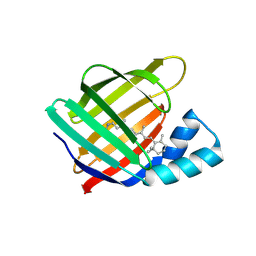
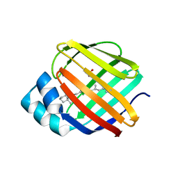
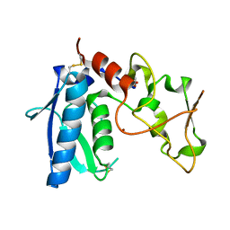
Crystal structure of the R132K:R111L:L121E:R59W-CRABPII mutant complexed with a synthetic ligand (merocyanin) at 2.60 angstrom resolution.
分子名称: (2E,4E,6E)-3-methyl-6-(1,3,3-trimethyl-1,3-dihydro-2H-indol-2-ylidene)hexa-2,4-dienal, 2-(N-MORPHOLINO)-ETHANESULFONIC ACID, Cellular retinoic acid-binding protein 2
著者Jia, X, Geiger, J.H.
登録日2008-11-30
公開日2009-11-10
最終更新日2023-09-06
実験手法X-RAY DIFFRACTION (2.6 Å)
主引用文献"Turn-on" protein fluorescence: in situ formation of cyanine dyes.
J.Am.Chem.Soc., 137, 2015
3CWK
DownloadVisualize
BU of 3cwk by Molmil
Crystal Structure of the R132K:Y134F:R111L:T54V:L121E Mutant of Cellular Retinoic Acid Binding Protein Type II in Complex with All-trans-Retinoic Acid at 1.57 Angstroms Resolution
分子名称: Cellular retinoic acid-binding protein 2, RETINOIC ACID, SULFATE ION
著者Vaezeslami, S, Geiger, J.H.
登録日2008-04-22
公開日2008-09-09
最終更新日2024-02-21
実験手法X-RAY DIFFRACTION (1.6 Å)
主引用文献Structural analysis of site-directed mutants of cellular retinoic acid-binding protein II addresses the relationship between structural integrity and ligand binding.
Acta Crystallogr.,Sect.D, 64, 2008
3FA9
DownloadVisualize
BU of 3fa9 by Molmil
Crystal structure of the apo R132K:Y134F:R111L:L121D mutant of cellular retinoic acid-binding protein II at 1.94 angstrom resolution
分子名称: ACETATE ION, Cellular retinoic acid-binding protein 2
著者Jia, X, Geiger, J.H.
登録日2008-11-16
公開日2009-10-27
最終更新日2023-12-27
実験手法X-RAY DIFFRACTION (1.94 Å)
主引用文献Crystal structures of apo cellular retinoic acid-binding protein II mutants: Structural integrity investigated through multiple site mutations
To be Published
3D96
DownloadVisualize
BU of 3d96 by Molmil
Crystal Structure of the R132K:Y134F Mutant of Apo-Cellular Retinoic Acid Binding Protein Type II at 1.71 Angstroms Resolution
分子名称: ACETATE ION, Cellular retinoic acid-binding protein 2
著者Vaezeslami, S, Geiger, J.H.
登録日2008-05-26
公開日2008-07-22
最終更新日2024-02-21
実験手法X-RAY DIFFRACTION (1.71 Å)
主引用文献Structural analysis of site-directed mutants of cellular retinoic acid-binding protein II addresses the relationship between structural integrity and ligand binding.
Acta Crystallogr.,Sect.D, 64, 2008
3FA8
DownloadVisualize
BU of 3fa8 by Molmil
Crystal structure of the apo R132K:Y134F:R111L:L121E mutant of cellular retinoic acid-binding protein II at 1.78 angstrom resolution
分子名称: Cellular retinoic acid-binding protein 2
著者Jia, X, Geiger, J.H.
登録日2008-11-16
公開日2009-10-27
最終更新日2023-09-06
実験手法X-RAY DIFFRACTION (1.78 Å)
主引用文献Crystal structures of apo cellular retinoic acid-binding protein II mutants: Structural integrity investigated through multiple site mutations
To be Published
3FEN
DownloadVisualize
BU of 3fen by Molmil
Crystal structure of the R132K:R111L:A32E mutant of cellular retinoic acid-binding protein II at 1.56 angstrom resolution
分子名称: CHLORIDE ION, Cellular retinoic acid-binding protein 2
著者Jia, X, Geiger, J.H.
登録日2008-11-30
公開日2009-11-10
最終更新日2023-09-06
実験手法X-RAY DIFFRACTION (1.56 Å)
主引用文献Crystal structures of apo cellular retinoic acid-binding protein II mutants: Structural integrity investigated through multiple site mutations
To be Published
3FA7
DownloadVisualize
BU of 3fa7 by Molmil
Crystal structure of the apo R132K:R111L:L121E:R59E mutant of cellular retinoic acid-binding protein II at 1.90 angstrom resolution
分子名称: 2-[3-(2-HYDROXY-1,1-DIHYDROXYMETHYL-ETHYLAMINO)-PROPYLAMINO]-2-HYDROXYMETHYL-PROPANE-1,3-DIOL, Cellular retinoic acid-binding protein 2
著者Jia, X, Geiger, J.H.
登録日2008-11-15
公開日2009-10-27
最終更新日2023-12-27
実験手法X-RAY DIFFRACTION (1.9 Å)
主引用文献Crystal structures of apo cellular retinoic acid-binding protein II mutants: Structural integrity investigated through multiple site mutations
To be Published
3FEK
DownloadVisualize
BU of 3fek by Molmil
Crystal structure of the R132K:Y134F:R111L:L121D:T54V mutant of cellular retinoic acid-binding protein II at 1.51 angstrom resolution
分子名称: ACETATE ION, Cellular retinoic acid-binding protein 2, DI(HYDROXYETHYL)ETHER
著者Jia, X, Geiger, J.H.
登録日2008-11-30
公開日2009-11-10
最終更新日2023-09-06
実験手法X-RAY DIFFRACTION (1.51 Å)
主引用文献Crystal structures of apo cellular retinoic acid-binding protein II mutants: Structural integrity investigated through multiple site mutations
To be Published
3EDG
DownloadVisualize
BU of 3edg by Molmil
Crystal structure of bone morphogenetic protein 1 protease domain
分子名称: ACETYL GROUP, Bone morphogenetic protein 1, ZINC ION
著者Mac Sweeney, A.
登録日2008-09-03
公開日2008-09-16
最終更新日2023-11-01
実験手法X-RAY DIFFRACTION (1.27 Å)
主引用文献Structural basis for the substrate specificity of bone morphogenetic protein 1/tolloid-like metalloproteases
To be Published
3D95
DownloadVisualize
BU of 3d95 by Molmil
Crystal Structure of the R132K:Y134F:R111L:L121E:T54V Mutant of Apo-Cellular Retinoic Acid Binding Protein Type II at 1.20 Angstroms Resolution
分子名称: Cellular retinoic acid-binding protein 2
著者Vaezeslami, S, Geiger, J.H.
登録日2008-05-26
公開日2008-07-22
最終更新日2024-02-21
実験手法X-RAY DIFFRACTION (1.2 Å)
主引用文献Structural analysis of site-directed mutants of cellular retinoic acid-binding protein II addresses the relationship between structural integrity and ligand binding.
Acta Crystallogr.,Sect.D, 64, 2008
3D97
DownloadVisualize
BU of 3d97 by Molmil
Crystal Structure of the R132K:R111L:L121E Mutant of Apo-Cellular Retinoic Acid Binding Protein Type II At 1.50 Angstroms Resolution
分子名称: 2-[3-(2-HYDROXY-1,1-DIHYDROXYMETHYL-ETHYLAMINO)-PROPYLAMINO]-2-HYDROXYMETHYL-PROPANE-1,3-DIOL, Cellular retinoic acid-binding protein 2, SODIUM ION
著者Vaezeslami, S, Geiger, J.H.
登録日2008-05-26
公開日2008-07-22
最終更新日2023-08-30
実験手法X-RAY DIFFRACTION (1.5 Å)
主引用文献Determining Crystal Structures of Proteins and Protein Complexes by X-Ray Crystallography: X-Ray Crystallographic Studies of the Mutants of Cellular Retinoic Acid Binding Protein Type II Toward Designing a Mimic of Rhodopsin.
Thesis

222415

件を2024-07-10に公開中

PDB statisticsPDBj update infoContact PDBjnumon