5YPE
 
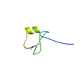 | | p62/SQSTM1 ZZ domain with Tyr-peptide | | 分子名称: | 78 kDa glucose-regulated protein,Sequestosome-1, ZINC ION | | 著者 | Kwon, D.H, Kim, L, Song, H.K. | | 登録日 | 2017-11-01 | | 公開日 | 2018-08-29 | | 最終更新日 | 2024-03-27 | | 実験手法 | X-RAY DIFFRACTION (2.851 Å) | | 主引用文献 | Insights into degradation mechanism of N-end rule substrates by p62/SQSTM1 autophagy adapter.
Nat Commun, 9, 2018
|
|
5YPA
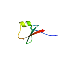 | | p62/SQSTM1 ZZ domain with Lys-peptide | | 分子名称: | 78 kDa glucose-regulated protein,Sequestosome-1, ZINC ION | | 著者 | Kwon, D.H, Kim, L, Song, H.K. | | 登録日 | 2017-11-01 | | 公開日 | 2018-08-29 | | 最終更新日 | 2024-03-27 | | 実験手法 | X-RAY DIFFRACTION (2.502 Å) | | 主引用文献 | Insights into degradation mechanism of N-end rule substrates by p62/SQSTM1 autophagy adapter.
Nat Commun, 9, 2018
|
|
5YP7
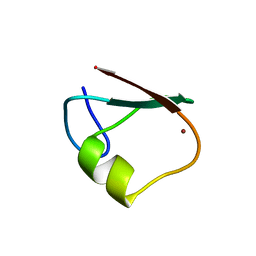 | | p62/SQSTM1 ZZ domain | | 分子名称: | Sequestosome-1, ZINC ION | | 著者 | Kwon, D.H, Kim, L, Song, H.K. | | 登録日 | 2017-11-01 | | 公開日 | 2018-08-29 | | 最終更新日 | 2024-03-27 | | 実験手法 | X-RAY DIFFRACTION (1.424 Å) | | 主引用文献 | Insights into degradation mechanism of N-end rule substrates by p62/SQSTM1 autophagy adapter.
Nat Commun, 9, 2018
|
|
6JHK
 | |
6K1D
 | |
6K1C
 | |
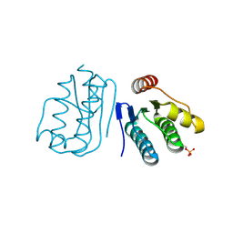
6K18
 | | Crystal structure of EXD2 exonuclease domain soaked in Mn | | 分子名称: | Exonuclease 3'-5' domain-containing protein 2, MANGANESE (II) ION | | 著者 | Park, J, Lee, C. | | 登録日 | 2019-05-10 | | 公開日 | 2019-05-22 | | 最終更新日 | 2023-11-22 | | 実験手法 | X-RAY DIFFRACTION (2.303 Å) | | 主引用文献 | The structure of human EXD2 reveals a chimeric 3' to 5' exonuclease domain that discriminates substrates via metal coordination.
Nucleic Acids Res., 47, 2019
|
|
6K1A
 | | Crystal structure of EXD2 exonuclease domain soaked in Mn and Mg | | 分子名称: | Exonuclease 3'-5' domain-containing protein 2, MAGNESIUM ION, MANGANESE (II) ION | | 著者 | Park, J, Lee, C. | | 登録日 | 2019-05-10 | | 公開日 | 2019-05-22 | | 最終更新日 | 2023-11-22 | | 実験手法 | X-RAY DIFFRACTION (2.602 Å) | | 主引用文献 | The structure of human EXD2 reveals a chimeric 3' to 5' exonuclease domain that discriminates substrates via metal coordination.
Nucleic Acids Res., 47, 2019
|
|
6K17
 | | Crystal structure of EXD2 exonuclease domain | | 分子名称: | Exonuclease 3'-5' domain-containing protein 2, SODIUM ION | | 著者 | Park, J, Lee, C. | | 登録日 | 2019-05-10 | | 公開日 | 2019-05-22 | | 最終更新日 | 2023-11-22 | | 実験手法 | X-RAY DIFFRACTION (1.602 Å) | | 主引用文献 | The structure of human EXD2 reveals a chimeric 3' to 5' exonuclease domain that discriminates substrates via metal coordination.
Nucleic Acids Res., 47, 2019
|
|
6K1E
 | |
6K1B
 | |
6K19
 | | Crystal structure of EXD2 exonuclease domain soaked in Mg | | 分子名称: | Exonuclease 3'-5' domain-containing protein 2, MAGNESIUM ION | | 著者 | Park, J, Lee, C. | | 登録日 | 2019-05-10 | | 公開日 | 2019-05-22 | | 最終更新日 | 2023-11-22 | | 実験手法 | X-RAY DIFFRACTION (2.202 Å) | | 主引用文献 | The structure of human EXD2 reveals a chimeric 3' to 5' exonuclease domain that discriminates substrates via metal coordination.
Nucleic Acids Res., 47, 2019
|
|


