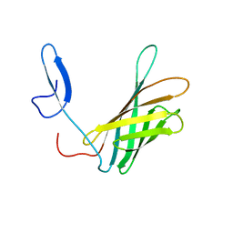
4AQP
 
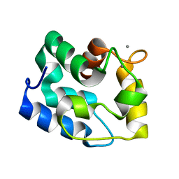
 | | The structure of the AXH domain of ataxin-1. | | 分子名称: | ATAXIN-1, DI(HYDROXYETHYL)ETHER, SODIUM ION | | 著者 | Rees, M, Chen, Y.W, de Chiara, C, Pastore, A. | | 登録日 | 2012-04-19 | | 公開日 | 2013-03-27 | | 最終更新日 | 2023-12-20 | | 実験手法 | X-RAY DIFFRACTION (2.452 Å) | | 主引用文献 | Self-Assembly and Conformational Heterogeneity of the Axh Domain of Ataxin-1: An Unusual Example of a Chameleon Fold
Biophys.J., 104, 2013
|
|
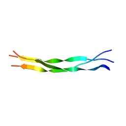
4APT
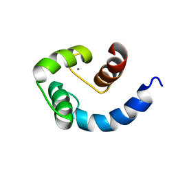
 | | The structure of the AXH domain of ataxin-1. | | 分子名称: | ATAXIN-1, SODIUM ION | | 著者 | Rees, M, Chen, Y.W, de Chiara, C, Pastore, A. | | 登録日 | 2012-04-05 | | 公開日 | 2013-03-27 | | 最終更新日 | 2023-12-20 | | 実験手法 | X-RAY DIFFRACTION (2.5 Å) | | 主引用文献 | Self-Assembly and Conformational Heterogeneity of the Axh Domain of Ataxin-1: An Unusual Example of a Chameleon Fold
Biophys.J., 104, 2013
|
|
2Y26
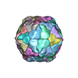 | | Transmission defective mutant of Grapevine Fanleaf virus | | 分子名称: | COAT PROTEIN | | 著者 | Schellenberger, P, Sauter, C, Lorber, B, Bron, P, Trapani, S, Bergdoll, M, Marmonier, A, Schmitt-Keichinger, C, Lemaire, O, Demangeat, G, Ritzenthaler, C. | | 登録日 | 2010-12-13 | | 公開日 | 2011-06-08 | | 最終更新日 | 2024-05-01 | | 実験手法 | X-RAY DIFFRACTION (2.7 Å) | | 主引用文献 | Structural Insights Into Viral Determinants of Nematode Mediated Grapevine Fanleaf Virus Transmission.
Plos Pathog., 7, 2011
|
|
1RK9
 | | Solution Structure of Human alpha-Parvalbumin (Minimized Average Structure) | | 分子名称: | CALCIUM ION, Parvalbumin alpha | | 著者 | Baig, I, Bertini, I, Del Bianco, C, Gupta, Y.K, Lee, Y.-M, Luchinat, C, Quattrone, A, Structural Proteomics in Europe (SPINE) | | 登録日 | 2003-11-21 | | 公開日 | 2004-06-08 | | 最終更新日 | 2024-05-22 | | 実験手法 | SOLUTION NMR | | 主引用文献 | Paramagnetism-based refinement strategy for the solution structure of human alpha-parvalbumin
Biochemistry, 43, 2004
|
|
1SW8
 | | Solution structure of the N-terminal domain of Human N60D calmodulin refined with paramagnetism based strategy | | 分子名称: | CALCIUM ION, Calmodulin | | 著者 | Bertini, I, Del Bianco, C, Gelis, I, Katsaros, N, Luchinat, C, Parigi, G, Peana, M, Provenzani, A, Zoroddu, M.A, Structural Proteomics in Europe (SPINE) | | 登録日 | 2004-03-30 | | 公開日 | 2004-04-06 | | 最終更新日 | 2024-05-22 | | 実験手法 | SOLUTION NMR | | 主引用文献 | Experimentally exploring the conformational space sampled by domain reorientation in calmodulin
Proc.Natl.Acad.Sci.USA, 101, 2004
|
|
5EUE
 | | S1P Lyase Bacterial Surrogate bound to N-(2-((4-methoxy-2,5-dimethylbenzyl)amino)-1-phenylethyl)-5-methylisoxazole-3-carboxamide | | 分子名称: | PHOSPHATE ION, Putative sphingosine-1-phosphate lyase, ~{N}-[(1~{S})-2-[(4-methoxy-2,5-dimethyl-phenyl)methylamino]-1-phenyl-ethyl]-5-methyl-1,2-oxazole-3-carboxamide | | 著者 | Argiriadi, M.A, Banach, D, Radziejewska, E, Marchie, S, DiMauro, J, Dinges, J, Dominguez, E, Hutchins, C, Judge, R.A, Queeney, K, Wallace, G, Harris, C.M. | | 登録日 | 2015-11-18 | | 公開日 | 2016-03-16 | | 最終更新日 | 2023-11-15 | | 実験手法 | X-RAY DIFFRACTION (2.83 Å) | | 主引用文献 | Creation of a S1P Lyase bacterial surrogate for structure-based drug design.
Bioorg.Med.Chem.Lett., 26, 2016
|
|
5EUD
 | | S1P Lyase Bacterial Surrogate bound to N-(1-(4-(3-hydroxyprop-1-yn-1-yl)phenyl)-2-((4-methoxy-2,5-dimethylbenzyl)amino)ethyl)-5-methylisoxazole-3-carboxamide | | 分子名称: | PHOSPHATE ION, Putative sphingosine-1-phosphate lyase, ~{N}-[(1~{S})-2-[(4-methoxy-2,5-dimethyl-phenyl)methylamino]-1-[4-(3-oxidanylprop-1-ynyl)phenyl]ethyl]-5-methyl-1,2-oxazole-3-carboxamide | | 著者 | Argiriadi, M.A, Banach, D, Radziejewska, E, Marchie, S, DiMauro, J, Dinges, J, Dominguez, E, Hutchins, C, Judge, R.A, Queeney, K, Wallace, G, Harris, C.M. | | 登録日 | 2015-11-18 | | 公開日 | 2016-03-16 | | 最終更新日 | 2023-11-15 | | 実験手法 | X-RAY DIFFRACTION (2.24 Å) | | 主引用文献 | Creation of a S1P Lyase bacterial surrogate for structure-based drug design.
Bioorg.Med.Chem.Lett., 26, 2016
|
|
1PIH
 | | THE THREE DIMENSIONAL STRUCTURE OF THE PARAMAGNETIC PROTEIN HIPIP I FROM E.HALOPHILA THROUGH NUCLEAR MAGNETIC RESONANCE | | 分子名称: | HIGH POTENTIAL IRON SULFUR PROTEIN, IRON/SULFUR CLUSTER | | 著者 | Banci, L, Bertini, I, Eltis, L.D, Felli, I, Kastrau, D.H.W, Luchinat, C, Piccioli, M, Pierattelli, R, Smith, M. | | 登録日 | 1994-08-03 | | 公開日 | 1994-12-20 | | 最終更新日 | 2024-05-22 | | 実験手法 | SOLUTION NMR | | 主引用文献 | The three-dimensional structure in solution of the paramagnetic high-potential iron-sulfur protein I from Ectothiorhodospira halophila through nuclear magnetic resonance.
Eur.J.Biochem., 225, 1994
|
|
1PIJ
 | | THE THREE DIMENSIONAL STRUCTURE OF THE PARAMAGNETIC PROTEIN HIPIP I FROM E.HALOPHILA THROUGH NUCLEAR MAGNETIC RESONANCE | | 分子名称: | HIGH POTENTIAL IRON SULFUR PROTEIN, IRON/SULFUR CLUSTER | | 著者 | Banci, L, Bertini, I, Eltis, L.D, Felli, I.C, Kastrau, D.H.W, Luchinat, C, Piccioli, M, Pierattelli, R, Smith, M. | | 登録日 | 1994-11-11 | | 公開日 | 1995-02-07 | | 最終更新日 | 2024-05-22 | | 実験手法 | SOLUTION NMR | | 主引用文献 | The three-dimensional structure in solution of the paramagnetic high-potential iron-sulfur protein I from Ectothiorhodospira halophila through nuclear magnetic resonance.
Eur.J.Biochem., 225, 1994
|
|
1RJV
 | | Solution Structure of Human alpha-Parvalbumin refined with a paramagnetism-based strategy | | 分子名称: | CALCIUM ION, Parvalbumin alpha | | 著者 | Baig, I, Bertini, I, Del Bianco, C, Gupta, Y.K, Lee, Y.M, Luchinat, C, Quattrone, A, Structural Proteomics in Europe (SPINE) | | 登録日 | 2003-11-20 | | 公開日 | 2004-05-25 | | 最終更新日 | 2024-05-22 | | 実験手法 | SOLUTION NMR | | 主引用文献 | Paramagnetism-Based Refinement Strategy for the Solution Structure of Human alpha-Parvalbumin.
Biochemistry, 43, 2004
|
|
3SHI
 | | Crystal structure of human MMP1 catalytic domain at 2.2 A resolution | | 分子名称: | CALCIUM ION, Interstitial collagenase, ZINC ION | | 著者 | Bertini, I, Calderone, V, Cerofolini, L, Fragai, M, Geraldes, C.F.G.C, Hermann, P, Luchinat, C, Parigi, G, Teixeira, J. | | 登録日 | 2011-06-16 | | 公開日 | 2011-09-21 | | 最終更新日 | 2023-09-13 | | 実験手法 | X-RAY DIFFRACTION (2.2 Å) | | 主引用文献 | The catalytic domain of MMP-1 studied through tagged lanthanides.
Febs Lett., 586, 2012
|
|
3NX7
 | | Crystal structure of the catalytic domain of human MMP12 complexed with the inhibitor N-Hydroxy-2-(N-(2-hydroxyethyl)4-methoxyphenylsulfonamido)acetamide | | 分子名称: | CALCIUM ION, Macrophage metalloelastase, N-hydroxy-N~2~-(2-hydroxyethyl)-N~2~-[(4-methoxyphenyl)sulfonyl]glycinamide, ... | | 著者 | Bertini, I, Calderone, V, Fragai, M, Giachetti, A, Loconte, M, Luchinat, C, Maletta, M, Nativi, C, Yeo, K.J. | | 登録日 | 2010-07-13 | | 公開日 | 2010-07-28 | | 最終更新日 | 2023-09-06 | | 実験手法 | X-RAY DIFFRACTION (1.8 Å) | | 主引用文献 | Exploring the subtleties of drug-receptor interactions: the case of matrix metalloproteinases
J.Am.Chem.Soc., 129, 2007
|
|
1TTX
 | | Solution Structure of human beta parvalbumin (oncomodulin) refined with a paramagnetism based strategy | | 分子名称: | CALCIUM ION, Oncomodulin | | 著者 | Babini, E, Bertini, I, Capozzi, F, Del Bianco, C, Hollender, D, Kiss, T, Luchinat, C, Quattrone, A, Structural Proteomics in Europe (SPINE) | | 登録日 | 2004-06-23 | | 公開日 | 2005-01-18 | | 最終更新日 | 2024-05-29 | | 実験手法 | SOLUTION NMR | | 主引用文献 | Solution Structure of Human beta-Parvalbumin and Structural Comparison with Its Paralog alpha-Parvalbumin and with Their Rat Orthologs(,)
Biochemistry, 43, 2004
|
|
2LKQ
 | | NMR structure of the lambda 5 22-45 peptide | | 分子名称: | Immunoglobulin lambda-like polypeptide 1 | | 著者 | Elantak, L, Espeli, M, Boned, A, Bornet, O, Breton, C, Feracci, M, Roche, P, Guerlesquin, F, Schiff, C. | | 登録日 | 2011-10-19 | | 公開日 | 2012-10-24 | | 最終更新日 | 2024-05-15 | | 実験手法 | SOLUTION NMR | | 主引用文献 | Structural Basis for Galectin-1-dependent Pre-B Cell Receptor (Pre-BCR) Activation.
J.Biol.Chem., 287, 2012
|
|
1PFD
 | | THE SOLUTION STRUCTURE OF HIGH PLANT PARSLEY [2FE-2S] FERREDOXIN, NMR, 18 STRUCTURES | | 分子名称: | FE2/S2 (INORGANIC) CLUSTER, FERREDOXIN | | 著者 | Im, S.-C, Liu, G, Luchinat, C, Sykes, A.G, Bertini, I. | | 登録日 | 1998-05-05 | | 公開日 | 1999-05-11 | | 最終更新日 | 2024-05-22 | | 実験手法 | SOLUTION NMR | | 主引用文献 | The solution structure of parsley [2Fe-2S]ferredoxin.
Eur.J.Biochem., 258, 1998
|
|
1BFY
 | | SOLUTION STRUCTURE OF REDUCED CLOSTRIDIUM PASTEURIANUM RUBREDOXIN, NMR, 20 STRUCTURES | | 分子名称: | FE (III) ION, RUBREDOXIN | | 著者 | Bertini, I, Kurtz Junior, D.M, Eidsness, M.K, Liu, G, Luchinat, C, Rosato, A, Scott, R.A. | | 登録日 | 1998-05-23 | | 公開日 | 1999-05-25 | | 最終更新日 | 2024-05-22 | | 実験手法 | SOLUTION NMR | | 主引用文献 | Solution Structure of Reduced Clostridium Pasteurianum Rubredoxin
J.Biol.Inorg.Chem., 3, 1998
|
|
5LHU
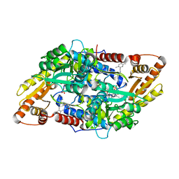
 | | ATP Phosphoribosyltransferase from Mycobacterium tuberculosis in complex with the allosteric inhibitor L-Histidine | | 分子名称: | ATP phosphoribosyltransferase, GLYCEROL, HISTIDINE, ... | | 著者 | de Chiara, C, Pisco, J.P, de Carvalho, L.P, Smerdon, S.J, Walker, P.A, Ogrodowicz, R. | | 登録日 | 2016-07-12 | | 公開日 | 2017-06-21 | | 最終更新日 | 2024-01-10 | | 実験手法 | X-RAY DIFFRACTION (2.02 Å) | | 主引用文献 | Uncoupling conformational states from activity in an allosteric enzyme.
Nat Commun, 8, 2017
|
|
1CKU
 | | AB INITIO SOLUTION AND REFINEMENT OF TWO HIGH POTENTIAL IRON PROTEIN STRUCTURES AT ATOMIC RESOLUTION | | 分子名称: | IRON/SULFUR CLUSTER, PROTEIN (HIPIP) | | 著者 | Parisini, E, Capozzi, F, Lubini, P, Lamzin, V, Luchinat, C, Sheldrick, G.M. | | 登録日 | 1999-04-24 | | 公開日 | 1999-05-13 | | 最終更新日 | 2023-12-27 | | 実験手法 | X-RAY DIFFRACTION (1.2 Å) | | 主引用文献 | Ab initio solution and refinement of two high-potential iron protein structures at atomic resolution.
Acta Crystallogr.,Sect.D, 55, 1999
|
|
7A70
 | | HEW lysozyme in complex with Ti(OH)4 | | 分子名称: | 1,2-ETHANEDIOL, CHLORIDE ION, Lysozyme, ... | | 著者 | Calderone, V, Gigli, L, Ravera, E, Luchinat, C. | | 登録日 | 2020-08-27 | | 公開日 | 2021-01-13 | | 最終更新日 | 2024-01-31 | | 実験手法 | X-RAY DIFFRACTION (1.8 Å) | | 主引用文献 | On the Mechanism of Bioinspired Formation of Inorganic Oxides: Structural Evidence of the Electrostatic Nature of the Interaction between a Mononuclear Inorganic Precursor and Lysozyme.
Biomolecules, 11, 2020
|
|
5LHT
 | | ATP Phosphoribosyltransferase from Mycobacterium tuberculosis in complex with the allosteric activator 3-(2-Thienyl)-L-alanine | | 分子名称: | ATP phosphoribosyltransferase, BETA(2-THIENYL)ALANINE, GLYCEROL, ... | | 著者 | de Chiara, C, Pisco, J.P, de Carvalho, L.P, Smerdon, S.J, Walker, P.A, Ogrodowicz, R. | | 登録日 | 2016-07-12 | | 公開日 | 2017-07-05 | | 最終更新日 | 2024-01-10 | | 実験手法 | X-RAY DIFFRACTION (2.0601 Å) | | 主引用文献 | Uncoupling conformational states from activity in an allosteric enzyme.
Nat Commun, 8, 2017
|
|
3ZYP
 | | Cellulose induced protein, Cip1 | | 分子名称: | 2-acetamido-2-deoxy-beta-D-glucopyranose, CALCIUM ION, CIP1, ... | | 著者 | Jacobson, F, Karkehabadi, S, Hansson, H, Goedegebuur, F, Wallace, L, Mitchinson, C, Piens, K, Stals, I, Sandgren, M. | | 登録日 | 2011-08-24 | | 公開日 | 2012-09-12 | | 最終更新日 | 2020-07-29 | | 実験手法 | X-RAY DIFFRACTION (1.5 Å) | | 主引用文献 | The Crystal Structure of the Core Domain of a Cellulose Induced Protein (Cip1) from Hypocrea Jecorina, at 1.5 A Resolution.
Plos One, 8, 2013
|
|
1UU5
 | | X-RAY CRYSTAL STRUCTURE OF THE CATALYTIC DOMAIN OF HUMICOLA GRISEA CEL12A SOAKED WITH CELLOTETRAOSE | | 分子名称: | ACETATE ION, ENDO-BETA-1,4-GLUCANASE, beta-D-glucopyranose-(1-4)-beta-D-glucopyranose-(1-3)-beta-D-glucopyranose-(1-4)-beta-D-glucopyranose | | 著者 | Berglund, G.I, Shaw, A, Stahlberg, J, Kenne, L, Driguez, T.H, Mitchinson, C, Sandgren, M. | | 登録日 | 2003-12-15 | | 公開日 | 2004-09-16 | | 最終更新日 | 2020-07-29 | | 実験手法 | X-RAY DIFFRACTION (1.67 Å) | | 主引用文献 | Crystal Complex Structures Reveal How Substrate is Bound in the -4 to the +2 Binding Sites of Humicola Grisea Cel12A
J.Mol.Biol., 342, 2004
|
|
2KRJ
 | | High-Resolution Solid-State NMR Structure of a 17.6 kDa Protein | | 分子名称: | COBALT (II) ION, Macrophage metalloelastase | | 著者 | Bertini, I, Bhaumik, A, De Pa pe, G, Griffin, R.G, Lelli, M, Lewandowski, J.R, Luchinat, C. | | 登録日 | 2009-12-18 | | 公開日 | 2010-02-23 | | 最終更新日 | 2024-05-01 | | 実験手法 | SOLID-STATE NMR | | 主引用文献 | High-resolution solid-state NMR structure of a 17.6 kDa protein.
J.Am.Chem.Soc., 132, 2010
|
|
2LFU
 | | The structure of a N. meningitides protein targeted for vaccine development | | 分子名称: | Gna2132 | | 著者 | Esposito, V, Musi, V, De Chiara, C, Kelly, G, Veggi, D, Pizza, M, Pastore, A. | | 登録日 | 2011-07-15 | | 公開日 | 2011-10-19 | | 最終更新日 | 2024-05-15 | | 実験手法 | SOLUTION NMR | | 主引用文献 | Structure of the C-terminal Domain of Neisseria Heparin Binding Antigen (NHBA), One of the Main Antigens of a Novel Vaccine against Neisseria meningitidis.
J.Biol.Chem., 286, 2011
|
|
2LLP
 | | Solution structure of a THP type 1 alpha 1 collagen fragment (772-786) | | 分子名称: | Collagen alpha-1(I) chain | | 著者 | Bertini, I, Fragai, M, Luchinat, C, Melikian, M, Toccafondi, M, Lauer, J.L, Fields, G.B, Structural Proteomics in Europe 2 (SPINE-2) | | 登録日 | 2011-11-15 | | 公開日 | 2012-05-30 | | 最終更新日 | 2024-05-01 | | 実験手法 | SOLUTION NMR | | 主引用文献 | Structural basis for matrix metalloproteinase 1-catalyzed collagenolysis.
J.Am.Chem.Soc., 134, 2012
|
|


