5NZR
 
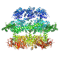
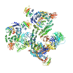 | | The structure of the COPI coat leaf | | 分子名称: | ADP-ribosylation factor 1, Coatomer subunit alpha, Coatomer subunit beta, ... | | 著者 | Dodonova, S.O, Aderhold, P, Kopp, J, Ganeva, I, Roehling, S, Hagen, W.J.H, Sinning, I, Wieland, F, Briggs, J.A.G. | | 登録日 | 2017-05-15 | | 公開日 | 2017-06-28 | | 最終更新日 | 2024-05-15 | | 実験手法 | ELECTRON MICROSCOPY (9.2 Å) | | 主引用文献 | 9 angstrom structure of the COPI coat reveals that the Arf1 GTPase occupies two contrasting molecular environments.
Elife, 6, 2017
|
|
5NZT
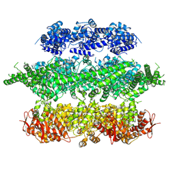
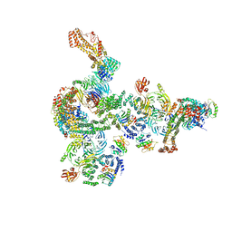 | | The structure of the COPI coat linkage I | | 分子名称: | ADP-ribosylation factor 1, Coatomer subunit alpha, Coatomer subunit beta, ... | | 著者 | Dodonova, S.O, Aderhold, P, Kopp, J, Ganeva, I, Roehling, S, Hagen, W.J.H, Sinning, I, Wieland, F, Briggs, J.A.G. | | 登録日 | 2017-05-15 | | 公開日 | 2017-06-28 | | 最終更新日 | 2024-05-15 | | 実験手法 | ELECTRON MICROSCOPY (17 Å) | | 主引用文献 | 9 angstrom structure of the COPI coat reveals that the Arf1 GTPase occupies two contrasting molecular environments.
Elife, 6, 2017
|
|
3MIX
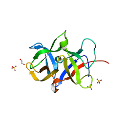
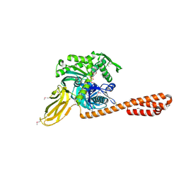
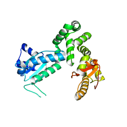 | | Crystal structure of the cytosolic domain of B. subtilis FlhA | | 分子名称: | Flagellar biosynthesis protein flhA | | 著者 | Bange, G, Kuemmerer, N, Bozkurt, G, Wild, K, Sinning, I. | | 登録日 | 2010-04-12 | | 公開日 | 2010-06-23 | | 最終更新日 | 2024-02-21 | | 実験手法 | X-RAY DIFFRACTION (2.3 Å) | | 主引用文献 | FlhA provides the adaptor for coordinated delivery of late flagella building blocks to the type III secretion system.
Proc.Natl.Acad.Sci.USA, 107, 2010
|
|
3KTV
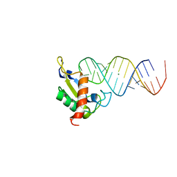
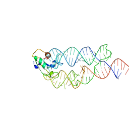 | | Crystal structure of the human SRP19/S-domain SRP RNA complex | | 分子名称: | MAGNESIUM ION, POTASSIUM ION, SRP RNA, ... | | 著者 | Wild, K, Bange, G, Bozkurt, G, Sinning, I. | | 登録日 | 2009-11-26 | | 公開日 | 2010-02-16 | | 最終更新日 | 2023-09-06 | | 実験手法 | X-RAY DIFFRACTION (3.8 Å) | | 主引用文献 | Structural insights into the assembly of the human and archaeal signal recognition particles.
Acta Crystallogr.,Sect.D, 66, 2010
|
|
3MAL
 | | Crystal structure of the SDF2-like protein from Arabidopsis thaliana | | 分子名称: | 1,2-ETHANEDIOL, SULFATE ION, Stromal cell-derived factor 2-like protein | | 著者 | Ravaud, S, Radzimanowski, J, Sinning, I. | | 登録日 | 2010-03-24 | | 公開日 | 2010-04-07 | | 最終更新日 | 2023-09-06 | | 実験手法 | X-RAY DIFFRACTION (1.95 Å) | | 主引用文献 | Arabidopsis stromal-derived Factor2 (SDF2) is a crucial target of the unfolded protein response in the endoplasmic reticulum.
J.Biol.Chem., 285, 2010
|
|
3BS6
 | |
5LNS
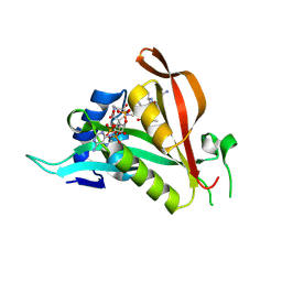
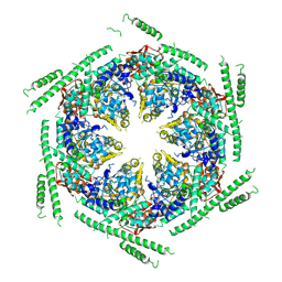
 | | Crystal structure of Arabidopsis thaliana Pdx1-R5P complex | | 分子名称: | PHOSPHATE ION, Pyridoxal 5'-phosphate synthase subunit PDX1.3, RIBULOSE-5-PHOSPHATE | | 著者 | Rodrigues, M.J, Windeisen, V, Zhang, Y, Guedez, G, Weber, S, Strohmeier, M, Hanes, J.W, Royant, A, Evans, G, Sinning, I, Ealick, S.E, Begley, T.P, Tews, I. | | 登録日 | 2016-08-06 | | 公開日 | 2017-01-18 | | 最終更新日 | 2024-10-16 | | 実験手法 | X-RAY DIFFRACTION (1.91 Å) | | 主引用文献 | Lysine relay mechanism coordinates intermediate transfer in vitamin B6 biosynthesis.
Nat. Chem. Biol., 13, 2017
|
|
5LNU
 | | Crystal structure of Arabidopsis thaliana Pdx1-I320 complex | | 分子名称: | (4~{S})-4-azanyl-5-oxidanyl-pent-1-en-3-one, PHOSPHATE ION, Pyridoxal 5'-phosphate synthase subunit PDX1.3, ... | | 著者 | Rodrigues, M.J, Windeisen, V, Zhang, Y, Guedez, G, Weber, S, Strohmeier, M, Hanes, J.W, Royant, A, Evans, G, Sinning, I, Ealick, S.E, Begley, T.P, Tews, I. | | 登録日 | 2016-08-06 | | 公開日 | 2017-01-18 | | 最終更新日 | 2024-10-16 | | 実験手法 | X-RAY DIFFRACTION (1.73 Å) | | 主引用文献 | Lysine relay mechanism coordinates intermediate transfer in vitamin B6 biosynthesis.
Nat. Chem. Biol., 13, 2017
|
|
5LNT
 | | Crystal structure of Arabidopsis thaliana Pdx1K166R-preI320 complex | | 分子名称: | PHOSPHATE ION, Pyridoxal 5'-phosphate synthase subunit PDX1.1, [(~{E},4~{S})-4-azanyl-3-oxidanylidene-pent-1-enyl] dihydrogen phosphate | | 著者 | Rodrigues, M.J, Windeisen, V, Zhang, Y, Guedez, G, Weber, S, Strohmeier, M, Hanes, J.W, Royant, A, Evans, G, Sinning, I, Ealick, S.E, Begley, T.P, Tews, I. | | 登録日 | 2016-08-06 | | 公開日 | 2017-01-18 | | 最終更新日 | 2024-10-16 | | 実験手法 | X-RAY DIFFRACTION (2.32 Å) | | 主引用文献 | Lysine relay mechanism coordinates intermediate transfer in vitamin B6 biosynthesis.
Nat. Chem. Biol., 13, 2017
|
|
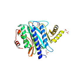
5LNR
 | | Crystal structure of Arabidopsis thaliana Pdx1-PLP complex | | 分子名称: | GLYCEROL, PYRIDOXAL-5'-PHOSPHATE, Pyridoxal 5'-phosphate synthase subunit PDX1.3 | | 著者 | Rodrigues, M.J, Windeisen, V, Zhang, Y, Guedez, G, Weber, S, Strohmeier, M, Hanes, J.W, Royant, A, Evans, G, Sinning, I, Ealick, S.E, Begley, T.P, Tews, I. | | 登録日 | 2016-08-06 | | 公開日 | 2017-01-18 | | 最終更新日 | 2025-04-09 | | 実験手法 | X-RAY DIFFRACTION (1.61 Å) | | 主引用文献 | Lysine relay mechanism coordinates intermediate transfer in vitamin B6 biosynthesis.
Nat. Chem. Biol., 13, 2017
|
|
5LNV
 | | Crystal structure of Arabidopsis thaliana Pdx1-I320 complex from multiple crystals | | 分子名称: | (4~{S})-4-azanyl-5-oxidanyl-pent-1-en-3-one, PHOSPHATE ION, Pyridoxal 5'-phosphate synthase subunit PDX1.3, ... | | 著者 | Rodrigues, M.J, Windeisen, V, Zhang, Y, Guedez, G, Weber, S, Strohmeier, M, Hanes, J.W, Royant, A, Evans, G, Sinning, I, Ealick, S.E, Begley, T.P, Tews, I. | | 登録日 | 2016-08-06 | | 公開日 | 2017-01-18 | | 最終更新日 | 2024-11-13 | | 実験手法 | X-RAY DIFFRACTION (2.24 Å) | | 主引用文献 | Lysine relay mechanism coordinates intermediate transfer in vitamin B6 biosynthesis.
Nat. Chem. Biol., 13, 2017
|
|
5LNW
 | | Crystal structure of Arabidopsis thaliana Pdx1-I320-G3P complex | | 分子名称: | 5-O-phosphono-beta-D-ribofuranose, GLYCEROL, Pyridoxal 5'-phosphate synthase subunit PDX1.3, ... | | 著者 | Rodrigues, M.J, Windeisen, V, Zhang, Y, Guedez, G, Weber, S, Strohmeier, M, Hanes, J.W, Royant, A, Evans, G, Sinning, I, Ealick, S.E, Begley, T.P, Tews, I. | | 登録日 | 2016-08-06 | | 公開日 | 2017-01-18 | | 最終更新日 | 2024-11-13 | | 実験手法 | X-RAY DIFFRACTION (1.9 Å) | | 主引用文献 | Lysine relay mechanism coordinates intermediate transfer in vitamin B6 biosynthesis.
Nat. Chem. Biol., 13, 2017
|
|
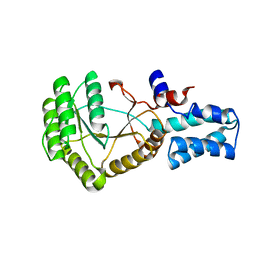
6TGX
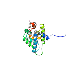
 | | Crystal structure of Arabidopsis thaliana NAA60 in complex with a bisubstrate analogue | | 分子名称: | Acyl-CoA N-acyltransferases (NAT) superfamily protein, CARBOXYMETHYL COENZYME *A, MET-VAL-ASN-ALA | | 著者 | Layer, D, Kopp, J, Lapouge, K, Sinning, I. | | 登録日 | 2019-11-18 | | 公開日 | 2020-06-24 | | 最終更新日 | 2024-11-13 | | 実験手法 | X-RAY DIFFRACTION (1.77 Å) | | 主引用文献 | The Arabidopsis N alpha -acetyltransferase NAA60 locates to the plasma membrane and is vital for the high salt stress response.
New Phytol., 228, 2020
|
|
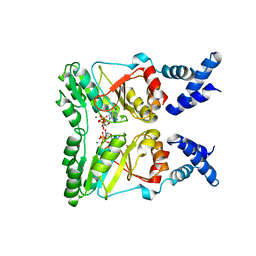
5MU7
 | | Crystal Structure of the beta/delta-COPI Core Complex | | 分子名称: | Coatomer subunit beta, Coatomer subunit delta-like protein | | 著者 | Kopp, J, Aderhold, P, Wieland, F, Sinning, I. | | 登録日 | 2017-01-12 | | 公開日 | 2017-06-28 | | 最終更新日 | 2024-05-08 | | 実験手法 | X-RAY DIFFRACTION (2.57 Å) | | 主引用文献 | 9 angstrom structure of the COPI coat reveals that the Arf1 GTPase occupies two contrasting molecular environments.
Elife, 6, 2017
|
|
1JID
 | |
3ZRJ
 | | Complex of ClpV N-domain with VipB peptide | | 分子名称: | 1,2-ETHANEDIOL, CLPB PROTEIN, VIPB | | 著者 | Lenherr, E.D, Kopp, J, Sinning, I. | | 登録日 | 2011-06-16 | | 公開日 | 2011-07-06 | | 最終更新日 | 2024-11-13 | | 実験手法 | X-RAY DIFFRACTION (1.94 Å) | | 主引用文献 | Molecular Basis for the Unique Role of the Aaa+ Chaperone Clpv in Type Vi Protein Secretion.
J.Biol.Chem., 286, 2011
|
|
4B9Q
 | |
3ZRI
 | |
4D2U
 | | Negative-stain electron microscopy of E. coli ClpB (BAP form bound to ClpP) | | 分子名称: | CHAPERONE PROTEIN CLPB | | 著者 | Carroni, M, Kummer, E, Oguchi, Y, Clare, D.K, Wendler, P, Sinning, I, Kopp, J, Mogk, A, Bukau, B, Saibil, H.R. | | 登録日 | 2014-05-13 | | 公開日 | 2014-06-04 | | 最終更新日 | 2024-11-13 | | 実験手法 | ELECTRON MICROSCOPY (17 Å) | | 主引用文献 | Head-to-Tail Interactions of the Coiled-Coil Domains Regulate Clpb Activity and Cooperation with Hsp70 in Protein Disaggregation.
Elife, 3, 2014
|
|
4D2Q
 | | Negative-stain electron microscopy of E. coli ClpB mutant E432A (BAP form bound to ClpP) | | 分子名称: | CLPB | | 著者 | Carroni, M, Kummer, E, Oguchi, Y, Clare, D.K, Wendler, P, Sinning, I, Kopp, J, Mogk, A, Bukau, B, Saibil, H.R. | | 登録日 | 2014-05-12 | | 公開日 | 2014-06-04 | | 最終更新日 | 2024-11-06 | | 実験手法 | ELECTRON MICROSCOPY (18 Å) | | 主引用文献 | Head-to-Tail Interactions of the Coiled-Coil Domains Regulate Clpb Activity and Cooperation with Hsp70 in Protein Disaggregation.
Elife, 3, 2014
|
|
4D2X
 | | Negative-stain electron microscopy of E. coli ClpB of Y503D hyperactive mutant (BAP form bound to ClpP) | | 分子名称: | CHAPERONE PROTEIN CLPB | | 著者 | Carroni, M, Kummer, E, Oguchi, Y, Clare, D.K, Wendler, P, Sinning, I, Kopp, J, Mogk, A, Bukau, B, Saibil, H.R. | | 登録日 | 2014-05-13 | | 公開日 | 2014-06-04 | | 最終更新日 | 2024-10-16 | | 実験手法 | ELECTRON MICROSCOPY (20 Å) | | 主引用文献 | Head-to-Tail Interactions of the Coiled-Coil Domains Regulate Clpb Activity and Cooperation with Hsp70 in Protein Disaggregation.
Elife, 3, 2014
|
|
2PX3
 | |
2QY9
 | |
2Q8K
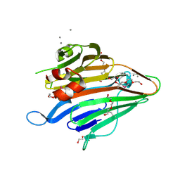
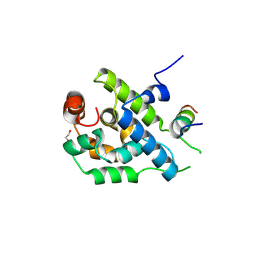
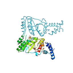
 | | The crystal structure of Ebp1 | | 分子名称: | GLYCEROL, Proliferation-associated protein 2G4, SULFATE ION | | 著者 | Kowalinski, E, Bange, G, Wild, K, Sinning, I. | | 登録日 | 2007-06-11 | | 公開日 | 2007-09-25 | | 最終更新日 | 2023-08-30 | | 実験手法 | X-RAY DIFFRACTION (1.6 Å) | | 主引用文献 | The crystal structure of Ebp1 reveals a methionine aminopeptidase fold as binding platform for multiple interactions.
Febs Lett., 581, 2007
|
|
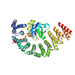
2PX0
 | | Crystal structure of FlhF complexed with GMPPNP/Mg(2+) | | 分子名称: | Flagellar biosynthesis protein flhF, MAGNESIUM ION, PHOSPHOAMINOPHOSPHONIC ACID-GUANYLATE ESTER | | 著者 | Bange, G, Wild, K, Sinning, I. | | 登録日 | 2007-05-14 | | 公開日 | 2007-09-25 | | 最終更新日 | 2024-02-21 | | 実験手法 | X-RAY DIFFRACTION (3 Å) | | 主引用文献 | The crystal structure of the third signal-recognition particle GTPase FlhF reveals a homodimer with bound GTP.
Proc.Natl.Acad.Sci.Usa, 104, 2007
|
|


