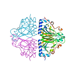
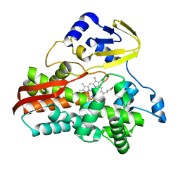
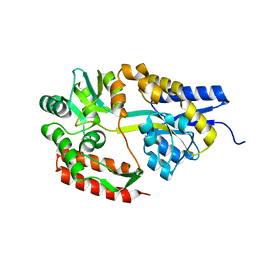
7WNK
 
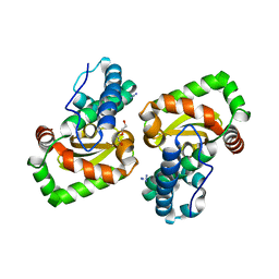 | | Crystal structure of a mutant Staphylococcus equorum manganese superoxide dismutase K38R and A121E | | 分子名称: | AZIDE ION, BROMIDE ION, DI(HYDROXYETHYL)ETHER, ... | | 著者 | Retnoningrum, D.S, Yoshida, H, Artarini, A.A, Ismaya, W.T. | | 登録日 | 2022-01-18 | | 公開日 | 2023-01-25 | | 最終更新日 | 2023-11-29 | | 実験手法 | X-RAY DIFFRACTION (1.4 Å) | | 主引用文献 | Introducing Intermolecular Interaction to Strengthen the Stability of MnSOD Dimer.
Appl.Biochem.Biotechnol., 195, 2023
|
|
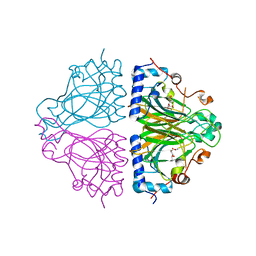
7WNL
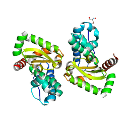 | | Crystal structure of a mutant Staphylococcus equorum manganese superoxide dismutase K38R and A121Y | | 分子名称: | 2-AMINO-2-HYDROXYMETHYL-PROPANE-1,3-DIOL, AZIDE ION, BROMIDE ION, ... | | 著者 | Retnoningrum, D.S, Yoshida, H, Artarini, A.A, Ismaya, W.T. | | 登録日 | 2022-01-18 | | 公開日 | 2023-01-25 | | 最終更新日 | 2023-11-29 | | 実験手法 | X-RAY DIFFRACTION (1.51 Å) | | 主引用文献 | Introducing Intermolecular Interaction to Strengthen the Stability of MnSOD Dimer.
Appl.Biochem.Biotechnol., 195, 2023
|
|
1RCL
 | |
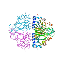
7W6W
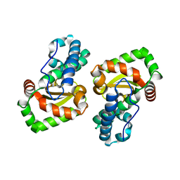 | | Crystal structure of a mutant Staphylococcus equorum manganese superoxide dismutase L169W | | 分子名称: | AZIDE ION, MANGANESE (II) ION, Superoxide dismutase | | 著者 | Retnoningrum, D.S, Yoshida, H, Artarini, A.A, Ismaya, W.T. | | 登録日 | 2021-12-02 | | 公開日 | 2022-12-07 | | 最終更新日 | 2023-11-29 | | 実験手法 | X-RAY DIFFRACTION (1.94 Å) | | 主引用文献 | Introducing Intermolecular Interaction to Strengthen the Stability of MnSOD Dimer.
Appl.Biochem.Biotechnol., 195, 2023
|
|
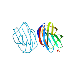
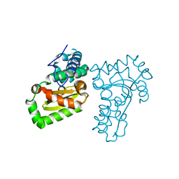
3VUS
 | | Escherichia coli PgaB N-terminal domain | | 分子名称: | ACETATE ION, MERCURY (II) ION, Poly-beta-1,6-N-acetyl-D-glucosamine N-deacetylase, ... | | 著者 | Nishiyama, T, Noguchi, H, Yoshida, H, Park, S.-Y, Tame, J.R.H. | | 登録日 | 2012-07-05 | | 公開日 | 2012-11-14 | | 最終更新日 | 2024-03-20 | | 実験手法 | X-RAY DIFFRACTION (1.65 Å) | | 主引用文献 | The structure of the deacetylase domain of Escherichia coli PgaB, an enzyme required for biofilm formation: a circularly permuted member of the carbohydrate esterase 4 family
Acta Crystallogr.,Sect.D, 69, 2013
|
|
3WUC
 | | X-ray crystal structure of Xenopus laevis galectin-Va | | 分子名称: | Galectin, MALONIC ACID, beta-D-galactopyranose-(1-4)-alpha-D-glucopyranose | | 著者 | Nonaka, Y, Yoshida, H, Kamitori, S, Nakamura, T. | | 登録日 | 2014-04-23 | | 公開日 | 2015-04-08 | | 最終更新日 | 2023-11-08 | | 実験手法 | X-RAY DIFFRACTION (1.6 Å) | | 主引用文献 | Crystal structure of a Xenopus laevis skin proto-type galectin, close to but distinct from galectin-1.
Glycobiology, 25, 2015
|
|
3WUD
 | | X-ray crystal structure of Xenopus laevis galectin-Ib | | 分子名称: | Galectin, SULFATE ION, beta-D-galactopyranose-(1-4)-alpha-D-glucopyranose | | 著者 | Nonaka, Y, Yoshida, H, Kamitori, S, Nakamura, T. | | 登録日 | 2014-04-23 | | 公開日 | 2015-04-08 | | 最終更新日 | 2023-11-08 | | 実験手法 | X-RAY DIFFRACTION (1.68 Å) | | 主引用文献 | Crystal structure of a Xenopus laevis skin proto-type galectin, close to but distinct from galectin-1.
Glycobiology, 25, 2015
|
|
5WQW
 | | X-ray structure of catalytic domain of autolysin from Clostridium perfringens | | 分子名称: | 1,2-ETHANEDIOL, N-acetylglucosaminidase | | 著者 | Tamai, E, Sekiya, H, Goda, E, Makihata, N, Maki, J, Yoshida, H, Kamitori, S. | | 登録日 | 2016-11-29 | | 公開日 | 2016-12-07 | | 最終更新日 | 2024-03-20 | | 実験手法 | X-RAY DIFFRACTION (1.76 Å) | | 主引用文献 | Structural and biochemical characterization of the Clostridium perfringens autolysin catalytic domain
FEBS Lett., 591, 2017
|
|
2ZYK
 | | Crystal structure of cyclo/maltodextrin-binding protein complexed with gamma-cyclodextrin | | 分子名称: | Cyclooctakis-(1-4)-(alpha-D-glucopyranose), Solute-binding protein | | 著者 | Tonozuka, T, Sogawa, A, Yamada, M, Matsumoto, N, Yoshida, H, Kamitori, S, Ichikawa, K, Mizuno, M, Nishikawa, A, Sakano, Y. | | 登録日 | 2009-01-26 | | 公開日 | 2009-02-10 | | 最終更新日 | 2024-04-03 | | 実験手法 | X-RAY DIFFRACTION (2.5 Å) | | 主引用文献 | Structural basis for cyclodextrin recognition by Thermoactinomyces vulgaris cyclo/maltodextrin-binding protein
Febs J., 274, 2007
|
|
2Z8G
 | | Aspergillus niger ATCC9642 isopullulanase complexed with isopanose | | 分子名称: | 2-acetamido-2-deoxy-beta-D-glucopyranose, Isopullulanase, alpha-D-glucopyranose-(1-4)-alpha-D-glucopyranose-(1-6)-beta-D-glucopyranose | | 著者 | Mizuno, M, Koide, A, Yamamura, A, Akeboshi, H, Yoshida, H, Kamitori, S, Sakano, Y, Nishikawa, A, Tonozuka, T. | | 登録日 | 2007-09-05 | | 公開日 | 2007-12-18 | | 最終更新日 | 2024-04-03 | | 実験手法 | X-RAY DIFFRACTION (1.7 Å) | | 主引用文献 | Crystal Structure of Aspergillus niger Isopullulanase, a Member of Glycoside Hydrolase Family 49
J.Mol.Biol., 376, 2008
|
|
3CW4
 | | Large c-terminal domain of influenza a virus RNA-dependent polymerase PB2 | | 分子名称: | Polymerase basic protein 2 | | 著者 | Kuzuhara, T, Kise, D, Yoshida, H, Horita, T, Murasaki, Y, Utsunomiya, H, Fujiki, H, Tsuge, H. | | 登録日 | 2008-04-21 | | 公開日 | 2009-01-13 | | 最終更新日 | 2024-03-20 | | 実験手法 | X-RAY DIFFRACTION (2.7 Å) | | 主引用文献 | Structural basis of the influenza A virus RNA polymerase PB2 RNA-binding domain containing the pathogenicity-determinant lysine 627 residue
J.Biol.Chem., 284, 2009
|
|
3W7T
 | | Escherichia coli K12 YgjK complexed with mannose | | 分子名称: | CALCIUM ION, MAGNESIUM ION, Uncharacterized protein YgjK, ... | | 著者 | Miyazaki, T, Kurakata, Y, Uechi, A, Yoshida, H, Kamitori, S, Sakano, Y, Nishikawa, A, Tonozuka, T. | | 登録日 | 2013-03-06 | | 公開日 | 2013-04-03 | | 最終更新日 | 2023-11-08 | | 実験手法 | X-RAY DIFFRACTION (1.5 Å) | | 主引用文献 | Structural insights into the substrate specificity and function of Escherichia coli K12 YgjK, a glucosidase belonging to the glycoside hydrolase family 63.
J.Mol.Biol., 381, 2008
|
|
3W7S
 | | Escherichia coli K12 YgjK complexed with glucose | | 分子名称: | CALCIUM ION, Uncharacterized protein YgjK, alpha-D-glucopyranose | | 著者 | Miyazaki, T, Kurakata, Y, Uechi, A, Yoshida, H, Kamitori, S, Sakano, Y, Nishikawa, A, Tonozuka, T. | | 登録日 | 2013-03-06 | | 公開日 | 2013-04-03 | | 最終更新日 | 2020-07-29 | | 実験手法 | X-RAY DIFFRACTION (1.9 Å) | | 主引用文献 | Structural insights into the substrate specificity and function of Escherichia coli K12 YgjK, a glucosidase belonging to the glycoside hydrolase family 63.
J.Mol.Biol., 381, 2008
|
|
3WW3
 | | X-ray structures of Cellulomonas parahominis L-ribose isomerase with no ligand | | 分子名称: | L-ribose isomerase, MANGANESE (II) ION | | 著者 | Terami, Y, Yoshida, H, Takata, G, Kamitori, S. | | 登録日 | 2014-06-13 | | 公開日 | 2015-04-29 | | 最終更新日 | 2024-05-29 | | 実験手法 | X-RAY DIFFRACTION (1.9 Å) | | 主引用文献 | Essentiality of tetramer formation of Cellulomonas parahominis L-ribose isomerase involved in novel L-ribose metabolic pathway.
Appl.Microbiol.Biotechnol., 99, 2015
|
|
3WW1
 | | X-ray structure of Cellulomonas parahominis L-ribose isomerase with L-ribose | | 分子名称: | L-ribose, L-ribose isomerase, MANGANESE (II) ION, ... | | 著者 | Terami, Y, Yoshida, H, Takata, G, Kamitori, S. | | 登録日 | 2014-06-13 | | 公開日 | 2015-04-29 | | 最終更新日 | 2024-05-29 | | 実験手法 | X-RAY DIFFRACTION (1.95 Å) | | 主引用文献 | Essentiality of tetramer formation of Cellulomonas parahominis L-ribose isomerase involved in novel L-ribose metabolic pathway.
Appl.Microbiol.Biotechnol., 99, 2015
|
|
3W7U
 | | Escherichia coli K12 YgjK complexed with galactose | | 分子名称: | CALCIUM ION, Uncharacterized protein YgjK, alpha-D-galactopyranose | | 著者 | Miyazaki, T, Kurakata, Y, Uechi, A, Yoshida, H, Kamitori, S, Sakano, Y, Nishikawa, A, Tonozuka, T. | | 登録日 | 2013-03-06 | | 公開日 | 2013-04-03 | | 最終更新日 | 2020-07-29 | | 実験手法 | X-RAY DIFFRACTION (1.99 Å) | | 主引用文献 | Structural insights into the substrate specificity and function of Escherichia coli K12 YgjK, a glucosidase belonging to the glycoside hydrolase family 63.
J.Mol.Biol., 381, 2008
|
|
3WW4
 | | X-ray structures of Cellulomonas parahominis L-ribose isomerase with L-allose | | 分子名称: | L-allose, L-ribose isomerase, MANGANESE (II) ION, ... | | 著者 | Terami, Y, Yoshida, H, Takata, G, Kamitori, S. | | 登録日 | 2014-06-13 | | 公開日 | 2015-04-29 | | 最終更新日 | 2024-05-29 | | 実験手法 | X-RAY DIFFRACTION (1.95 Å) | | 主引用文献 | Essentiality of tetramer formation of Cellulomonas parahominis L-ribose isomerase involved in novel L-ribose metabolic pathway.
Appl.Microbiol.Biotechnol., 99, 2015
|
|
3WW2
 | | X-ray structures of Cellulomonas parahominis L-ribose isomerase with L-psicose | | 分子名称: | L-psicose, L-ribose isomerase, MANGANESE (II) ION, ... | | 著者 | Terami, Y, Yoshida, H, Takata, G, Kamitori, S. | | 登録日 | 2014-06-13 | | 公開日 | 2015-04-29 | | 最終更新日 | 2024-05-29 | | 実験手法 | X-RAY DIFFRACTION (2 Å) | | 主引用文献 | Essentiality of tetramer formation of Cellulomonas parahominis L-ribose isomerase involved in novel L-ribose metabolic pathway.
Appl.Microbiol.Biotechnol., 99, 2015
|
|
3B4X
 | |
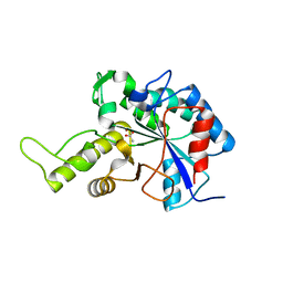
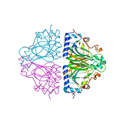
5X2J
 | | Crystal structure of a recombinant hybrid manganese superoxide dismutase from Staphylococcus equorum and Staphylococcus saprophyticus | | 分子名称: | MANGANESE (II) ION, manganese superoxide dismutase | | 著者 | Retnoningrum, D.S, Yoshida, H, Arumsari, S, Kamitori, S, Ismaya, W.T. | | 登録日 | 2017-01-31 | | 公開日 | 2018-01-31 | | 最終更新日 | 2023-11-22 | | 実験手法 | X-RAY DIFFRACTION (1.4 Å) | | 主引用文献 | The first crystal structure of manganese superoxide dismutase from the genus Staphylococcus
Acta Crystallogr F Struct Biol Commun, 74, 2018
|
|
2ZKN
 | |
5ZQT
 | | Crystal structure of Oryza sativa hexokinase 6 | | 分子名称: | Hexokinase-6, MAGNESIUM ION, PHOSPHOAMINOPHOSPHONIC ACID-ADENYLATE ESTER, ... | | 著者 | Matsudaira, K, Mochizuki, S, Yoshida, H, Kamitori, S, Akimitsu, K. | | 登録日 | 2018-04-20 | | 公開日 | 2019-04-24 | | 最終更新日 | 2023-11-22 | | 実験手法 | X-RAY DIFFRACTION (2.84 Å) | | 主引用文献 | Crystal structure of Oryza sativa hexokinase 6
To Be Published
|
|
2EIS
 | | X-ray structure of acyl-CoA hydrolase-like protein, TT1379, from Thermus thermophilus HB8 | | 分子名称: | COENZYME A, Hypothetical protein TTHB207 | | 著者 | Kamitori, S, Yoshida, H, Satoh, S, Iino, H, Ebihara, A, Chen, L, Fu, Z.-Q, Chrzas, J, Wang, B.-C, Yokoyama, S, Kuramitsu, S, RIKEN Structural Genomics/Proteomics Initiative (RSGI) | | 登録日 | 2007-03-13 | | 公開日 | 2008-03-18 | | 最終更新日 | 2011-07-13 | | 実験手法 | X-RAY DIFFRACTION (2.1 Å) | | 主引用文献 | X-ray structure of acyl-CoA hydrolase-like protein, TT1379, from Thermus thermophilus HB8
To be Published
|
|
2ZYO
 | | Crystal structure of cyclo/maltodextrin-binding protein complexed with maltotetraose | | 分子名称: | alpha-D-glucopyranose-(1-4)-alpha-D-glucopyranose, solute-binding protein | | 著者 | Matsumoto, M, Yamada, M, Kurakata, Y, Yoshida, H, Kamitori, S, Nishikawa, A, Tonozuka, T. | | 登録日 | 2009-01-27 | | 公開日 | 2009-03-31 | | 最終更新日 | 2023-11-01 | | 実験手法 | X-RAY DIFFRACTION (1.55 Å) | | 主引用文献 | Crystal structures of open and closed forms of cyclo/maltodextrin-binding protein
Febs J., 276, 2009
|
|
2ZYM
 | | Crystal structure of cyclo/maltodextrin-binding protein complexed with alpha-cyclodextrin | | 分子名称: | Cyclohexakis-(1-4)-(alpha-D-glucopyranose), Solute-binding protein | | 著者 | Matsumoto, M, Yamada, M, Kurakata, Y, Yoshida, H, Kamitori, S, Nishikawa, A, Tonozuka, T. | | 登録日 | 2009-01-27 | | 公開日 | 2009-03-31 | | 最終更新日 | 2023-11-01 | | 実験手法 | X-RAY DIFFRACTION (1.8 Å) | | 主引用文献 | Crystal structures of open and closed forms of cyclo/maltodextrin-binding protein
Febs J., 276, 2009
|
|


