4PP0
 
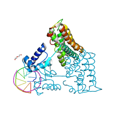
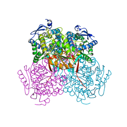
 | | Structure of the PBP NocT-M117N in complex with pyronopaline | | 分子名称: | 1,2-ETHANEDIOL, 1-[(1S)-4-carbamimidamido-1-carboxybutyl]-5-oxo-D-proline, DI(HYDROXYETHYL)ETHER, ... | | 著者 | Morera, S, Vigouroux, A. | | 登録日 | 2014-02-26 | | 公開日 | 2014-10-22 | | 最終更新日 | 2023-09-20 | | 実験手法 | X-RAY DIFFRACTION (1.57 Å) | | 主引用文献 | Agrobacterium uses a unique ligand-binding mode for trapping opines and acquiring a competitive advantage in the niche construction on plant host.
Plos Pathog., 10, 2014
|
|
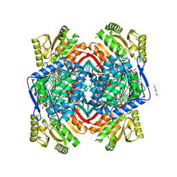
6ZK1
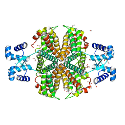
 | | Plant nucleoside hydrolase - ZmNRh2b enzyme | | 分子名称: | 1,2-ETHANEDIOL, CALCIUM ION, CHLORIDE ION, ... | | 著者 | Morera, S, Vigouroux, A, Kopecny, D. | | 登録日 | 2020-06-29 | | 公開日 | 2022-01-12 | | 最終更新日 | 2024-01-31 | | 実験手法 | X-RAY DIFFRACTION (1.99 Å) | | 主引用文献 | Plant nucleoside N-ribohydrolases: riboside binding and role in nitrogen storage mobilization.
Plant J., 2023
|
|
6ZK2
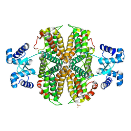
 | | Plant nucleoside hydrolase - ZmNRh2b in complex with forodesine | | 分子名称: | 1,2-ETHANEDIOL, 1,4-DIDEOXY-4-AZA-1-(S)-(9-DEAZAHYPOXANTHIN-9-YL)-D-RIBITOL, CALCIUM ION, ... | | 著者 | Morera, S, Vigouroux, A, Kopecny, D. | | 登録日 | 2020-06-29 | | 公開日 | 2022-01-12 | | 最終更新日 | 2024-01-31 | | 実験手法 | X-RAY DIFFRACTION (2.2 Å) | | 主引用文献 | Plant nucleoside N-ribohydrolases: riboside binding and role in nitrogen storage mobilization.
Plant J., 2023
|
|
6ZK3
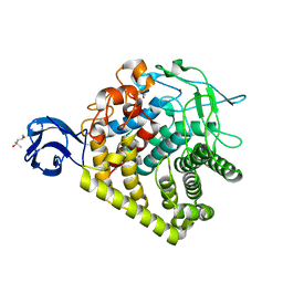
 | | Plant nucleoside hydrolase - ZmNRh2b in complex with ribose | | 分子名称: | 1,2-ETHANEDIOL, CALCIUM ION, DI(HYDROXYETHYL)ETHER, ... | | 著者 | Morera, S, Vigouroux, A, Kopecny, D. | | 登録日 | 2020-06-29 | | 公開日 | 2022-01-12 | | 最終更新日 | 2024-01-31 | | 実験手法 | X-RAY DIFFRACTION (1.75 Å) | | 主引用文献 | Plant nucleoside N-ribohydrolases: riboside binding and role in nitrogen storage mobilization.
Plant J., 2023
|
|
6ZK4
 | | Plant nucleoside hydrolase - ZmNRh2b with a bound adenine | | 分子名称: | 1,2-ETHANEDIOL, ADENINE, CALCIUM ION, ... | | 著者 | Morera, S, Vigouroux, A, Kopecny, D. | | 登録日 | 2020-06-29 | | 公開日 | 2022-01-12 | | 最終更新日 | 2024-01-31 | | 実験手法 | X-RAY DIFFRACTION (1.95 Å) | | 主引用文献 | Plant nucleoside N-ribohydrolases: riboside binding and role in nitrogen storage mobilization.
Plant J., 2023
|
|
6ZK5
 | | Plant nucleoside hydrolase - ZmNRh3 enzyme in complex with forodesine | | 分子名称: | 1,2-ETHANEDIOL, 1,4-DIDEOXY-4-AZA-1-(S)-(9-DEAZAHYPOXANTHIN-9-YL)-D-RIBITOL, CALCIUM ION, ... | | 著者 | Morera, S, Vigouroux, A, Kopecny, D. | | 登録日 | 2020-06-29 | | 公開日 | 2022-01-12 | | 最終更新日 | 2024-01-31 | | 実験手法 | X-RAY DIFFRACTION (1.9 Å) | | 主引用文献 | Plant nucleoside N-ribohydrolases: riboside binding and role in nitrogen storage mobilization.
Plant J., 2023
|
|
6ZA0
 | | Structure of the transcriptional repressor Atu1419 (VanR) in complex with a fortuitous citrate from agrobacterium fabrum (P21212 space group) | | 分子名称: | 1,2-ETHANEDIOL, CITRIC ACID, DI(HYDROXYETHYL)ETHER, ... | | 著者 | Morera, S, Vigouroux, A, Legrand, P. | | 登録日 | 2020-06-04 | | 公開日 | 2020-12-02 | | 最終更新日 | 2024-01-24 | | 実験手法 | X-RAY DIFFRACTION (1.65 Å) | | 主引用文献 | Characterization of the first tetrameric transcription factor of the GntR superfamily with allosteric regulation from the bacterial pathogen Agrobacterium fabrum.
Nucleic Acids Res., 49, 2021
|
|
6ZA3
 | | Structure of the transcriptional repressor Atu1419 (VanR) from agrobacterium fabrum in complex a palindromic DNA (C2221 space group) | | 分子名称: | 1,2-ETHANEDIOL, ACETATE ION, CHLORIDE ION, ... | | 著者 | Morera, S, Vigouroux, A, Legrand, P. | | 登録日 | 2020-06-04 | | 公開日 | 2020-12-02 | | 最終更新日 | 2024-01-24 | | 実験手法 | X-RAY DIFFRACTION (2.05 Å) | | 主引用文献 | Characterization of the first tetrameric transcription factor of the GntR superfamily with allosteric regulation from the bacterial pathogen Agrobacterium fabrum.
Nucleic Acids Res., 49, 2021
|
|
6ZAB
 | | Structure of the transcriptional repressor Atu1419 (VanR) from agrobacterium fabrum in complex a palindromic DNA (P6422 space group) | | 分子名称: | 1,2-ETHANEDIOL, CITRIC ACID, DI(HYDROXYETHYL)ETHER, ... | | 著者 | Morera, S, Naretto, A, Vigouroux, A, Legrand, P. | | 登録日 | 2020-06-05 | | 公開日 | 2020-12-02 | | 最終更新日 | 2024-01-24 | | 実験手法 | X-RAY DIFFRACTION (2.8 Å) | | 主引用文献 | Characterization of the first tetrameric transcription factor of the GntR superfamily with allosteric regulation from the bacterial pathogen Agrobacterium fabrum.
Nucleic Acids Res., 49, 2021
|
|
6Z74
 | |
6ZA7
 | | Structure of the apo transcriptional repressor Atu1419 (VanR) from agrobacterium fabrum | | 分子名称: | DI(HYDROXYETHYL)ETHER, GLYCEROL, SULFATE ION, ... | | 著者 | Morera, S, Vigouroux, A, Legrand, P. | | 登録日 | 2020-06-04 | | 公開日 | 2020-12-02 | | 最終更新日 | 2024-01-24 | | 実験手法 | X-RAY DIFFRACTION (2.34 Å) | | 主引用文献 | Characterization of the first tetrameric transcription factor of the GntR superfamily with allosteric regulation from the bacterial pathogen Agrobacterium fabrum.
Nucleic Acids Res., 49, 2021
|
|
3GZK
 | | Structure of A. Acidocaldarius Cellulase CelA | | 分子名称: | (4S)-2-METHYL-2,4-PENTANEDIOL, CALCIUM ION, Cellulase, ... | | 著者 | Morera, S, Eckert, K, Vigouroux, A. | | 登録日 | 2009-04-07 | | 公開日 | 2009-10-13 | | 最終更新日 | 2023-11-01 | | 実験手法 | X-RAY DIFFRACTION (1.8 Å) | | 主引用文献 | Crystal structures of A. acidocaldarius endoglucanase Cel9A in complex with cello-oligosaccharides: strong -1 and -2 subsites mimic cellobiohydrolase activity
J.Mol.Biol., 394, 2009
|
|
4PXN
 | |
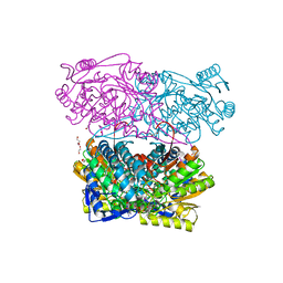
4PZ2
 | | Structure of Zm ALDH2-6 (RF2F) in complex with NAD | | 分子名称: | 1,2-ETHANEDIOL, DI(HYDROXYETHYL)ETHER, NICOTINAMIDE-ADENINE-DINUCLEOTIDE, ... | | 著者 | Morera, S, Vigouroux, A, Kopecny, D. | | 登録日 | 2014-03-28 | | 公開日 | 2015-03-18 | | 最終更新日 | 2023-09-20 | | 実験手法 | X-RAY DIFFRACTION (2.4 Å) | | 主引用文献 | Role and structural characterization of plant aldehyde dehydrogenases from family 2 and family 7.
Biochem.J., 468, 2015
|
|
4PXL
 | | Structure of Zm ALDH2-3 (RF2C) in complex with NAD | | 分子名称: | 1,2-ETHANEDIOL, CALCIUM ION, Cytosolic aldehyde dehydrogenase RF2C, ... | | 著者 | Morera, S, Vigouroux, A, Kopecny, D. | | 登録日 | 2014-03-24 | | 公開日 | 2015-03-18 | | 最終更新日 | 2024-11-27 | | 実験手法 | X-RAY DIFFRACTION (2.25 Å) | | 主引用文献 | Role and structural characterization of plant aldehyde dehydrogenases from family 2 and family 7.
Biochem.J., 468, 2015
|
|
5CFE
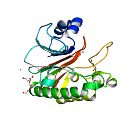 | | Bacillus subtilis AP endonuclease ExoA | | 分子名称: | CALCIUM ION, DI(HYDROXYETHYL)ETHER, Exodeoxyribonuclease | | 著者 | Morera, S, Vigouroux, A. | | 登録日 | 2015-07-08 | | 公開日 | 2016-07-06 | | 最終更新日 | 2024-01-10 | | 実験手法 | X-RAY DIFFRACTION (1.84 Å) | | 主引用文献 | Structural comparison of AP endonucleases from the exonuclease III family reveals new amino acid residues in human AP endonuclease 1 that are involved in incision of damaged DNA.
Biochimie, 128-129, 2016
|
|
5CFG
 | | C2 crystal form of APE1 with Mg2+ | | 分子名称: | DNA-(apurinic or apyrimidinic site) lyase, MAGNESIUM ION | | 著者 | Morera, S, Vigouroux, A. | | 登録日 | 2015-07-08 | | 公開日 | 2016-07-06 | | 最終更新日 | 2024-11-13 | | 実験手法 | X-RAY DIFFRACTION (1.8 Å) | | 主引用文献 | Structural comparison of AP endonucleases from the exonuclease III family reveals new amino acid residues in human AP endonuclease 1 that are involved in incision of damaged DNA.
Biochimie, 128-129, 2016
|
|
1NPK
 | | REFINED X-RAY STRUCTURE OF DICTYOSTELIUM NUCLEOSIDE DIPHOSPHATE KINASE AT 1,8 ANGSTROMS RESOLUTION | | 分子名称: | NUCLEOSIDE DIPHOSPHATE KINASE | | 著者 | Janin, J, Morera, S, Lebras, G, Lascu, I, Veron, M. | | 登録日 | 1994-07-27 | | 公開日 | 1994-11-01 | | 最終更新日 | 2024-02-14 | | 実験手法 | X-RAY DIFFRACTION (1.8 Å) | | 主引用文献 | Refined X-ray structure of Dictyostelium discoideum nucleoside diphosphate kinase at 1.8 A resolution.
J.Mol.Biol., 243, 1994
|
|
1MN7
 | | NDP kinase mutant (H122G;N119S;F64W) in complex with aBAZTTP | | 分子名称: | 3'-AZIDO-3'-DEOXY-THYMIDINE-5'-ALPHA BORANO TRIPHOSPHATE, MAGNESIUM ION, NDP kinase | | 著者 | gallois-montbrun, s, schneider, b, chen, y, giacomoni-fernandes, v, mulard, l, morera, s, janin, j, deville-bonne, d, veron, m. | | 登録日 | 2002-09-05 | | 公開日 | 2002-10-02 | | 最終更新日 | 2024-05-29 | | 実験手法 | X-RAY DIFFRACTION (2.15 Å) | | 主引用文献 | Improving nucleoside diphosphate kinase for antiviral nucleotide analogs activation
J.BIOL.CHEM., 277, 2002
|
|
1M5R
 | | Ternary complex of T4 phage BGT with UDP and a 13 mer DNA duplex | | 分子名称: | (4S)-2-METHYL-2,4-PENTANEDIOL, 2-AMINO-2-HYDROXYMETHYL-PROPANE-1,3-DIOL, 5'-D(*CP*TP*AP*TP*CP*TP*GP*AP*GP*TP*AP*TP*C)-3', ... | | 著者 | Lariviere, L, Morera, S. | | 登録日 | 2002-07-10 | | 公開日 | 2002-12-11 | | 最終更新日 | 2024-02-14 | | 実験手法 | X-RAY DIFFRACTION (1.8 Å) | | 主引用文献 | A Base-flipping mechanism for the T4 phage beta-glucosyltransferase and
identification of a transition state analog
J.Mol.Biol., 324, 2002
|
|
9FW6
 | | A ternary complex of plant adenosine kinase 1 from moss Physcomitrella patens (PpADK1) with adenosine and ADP | | 分子名称: | 1,2-ETHANEDIOL, ADENOSINE, ADENOSINE-5'-DIPHOSPHATE, ... | | 著者 | Kopecny, D, Vigouroux, A, Morera, S. | | 登録日 | 2024-06-28 | | 公開日 | 2025-04-09 | | 実験手法 | X-RAY DIFFRACTION (1.73 Å) | | 主引用文献 | A monomer-dimer switch modulates the activity of plant adenosine kinase.
J.Exp.Bot., 2025
|
|
2LSU
 | | The NMR high resolution structure of yeast Tah1 in a free form | | 分子名称: | TPR repeat-containing protein associated with Hsp90 | | 著者 | Back, R, Dominguez, C, Rothe, B, Bobo, C, Beaufils, C, Morera, S, Meyer, P, Charpentier, B, Branlant, C, Allain, F, Manival, X. | | 登録日 | 2012-05-07 | | 公開日 | 2013-05-22 | | 最終更新日 | 2024-05-15 | | 実験手法 | SOLUTION NMR | | 主引用文献 | High-Resolution Structural Analysis Shows How Tah1 Tethers Hsp90 to the R2TP Complex.
Structure, 21, 2013
|
|
2LSV
 | | The NMR high resolution structure of yeast Tah1 in complex with the Hsp90 C-terminal tail | | 分子名称: | ATP-dependent molecular chaperone HSP82, TPR repeat-containing protein associated with Hsp90 | | 著者 | Back, R, Dominguez, C, Rothe, B, Bobo, C, Beaufils, C, Morera, S, Meyer, P, Charpentier, B, Branlant, C, Allain, F, Manival, X. | | 登録日 | 2012-05-07 | | 公開日 | 2013-05-22 | | 最終更新日 | 2024-05-15 | | 実験手法 | SOLUTION NMR | | 主引用文献 | High-Resolution Structural Analysis Shows How Tah1 Tethers Hsp90 to the R2TP Complex.
Structure, 21, 2013
|
|
2QMH
 | | structure of V267F mutant HprK/P | | 分子名称: | HPr kinase/phosphorylase | | 著者 | Chaptal, V, Vincent, F, Gueguen-Chaignon, V, Poncet, S, Deutscher, J, Nessler, S, Morera, S. | | 登録日 | 2007-07-16 | | 公開日 | 2007-09-18 | | 最終更新日 | 2023-08-30 | | 実験手法 | X-RAY DIFFRACTION (2.6 Å) | | 主引用文献 | Structural Analysis of the Bacterial HPr Kinase/Phosphorylase V267F Mutant Gives Insights into the Allosteric Regulation Mechanism of This Bifunctional Enzyme.
J.Biol.Chem., 282, 2007
|
|
1G6Y
 | | CRYSTAL STRUCTURE OF THE GLOBULAR REGION OF THE PRION PROTEIN URE2 FROM YEAST SACCHAROMYCES CEREVISIAE | | 分子名称: | URE2 PROTEIN | | 著者 | Bousset, L, Belrhali, H, Janin, J, Melki, R, Morera, S. | | 登録日 | 2000-11-08 | | 公開日 | 2001-02-21 | | 最終更新日 | 2024-02-07 | | 実験手法 | X-RAY DIFFRACTION (2.8 Å) | | 主引用文献 | Structure of the globular region of the prion protein Ure2 from the yeast Saccharomyces cerevisiae.
Structure, 9, 2001
|
|


