5MNY
 
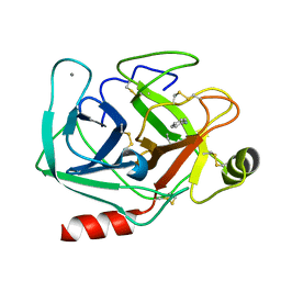 | | Neutron structure of cationic trypsin in complex with aniline | | 分子名称: | CALCIUM ION, Cationic trypsin, phenylazanium | | 著者 | Schiebel, J, Schrader, T.E, Ostermann, A, Heine, A, Klebe, G. | | 登録日 | 2016-12-13 | | 公開日 | 2017-05-24 | | 最終更新日 | 2024-10-16 | | 実験手法 | NEUTRON DIFFRACTION (1.43 Å) | | 主引用文献 | Charges Shift Protonation: Neutron Diffraction Reveals that Aniline and 2-Aminopyridine Become Protonated Upon Binding to Trypsin.
Angew. Chem. Int. Ed. Engl., 56, 2017
|
|
5MO1
 
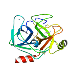 | | Neutron structure of cationic trypsin in complex with benzylamine | | 分子名称: | (phenylmethyl)azanium, CALCIUM ION, Cationic trypsin | | 著者 | Schiebel, J, Schrader, T.E, Ostermann, A, Heine, A, Klebe, G. | | 登録日 | 2016-12-13 | | 公開日 | 2018-02-28 | | 最終更新日 | 2024-01-17 | | 実験手法 | NEUTRON DIFFRACTION (1.491 Å) | | 主引用文献 | Neutron structure of cationic trypsin in complex with benzylamine
to be published
|
|
5MOR
 
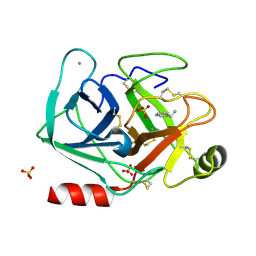 | | Joint X-ray/neutron structure of cationic trypsin in complex with benzylamine | | 分子名称: | (phenylmethyl)azanium, CALCIUM ION, Cationic trypsin, ... | | 著者 | Schiebel, J, Schrader, T.E, Ostermann, A, Heine, A, Klebe, G. | | 登録日 | 2016-12-14 | | 公開日 | 2018-02-28 | | 最終更新日 | 2024-10-23 | | 実験手法 | NEUTRON DIFFRACTION (0.98 Å), X-RAY DIFFRACTION | | 主引用文献 | Joint X-ray/neutron structure of cationic trypsin in complex with benzylamine
to be published
|
|
5MOO
 
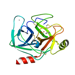 | | Joint X-ray/neutron structure of cationic trypsin in complex with aniline | | 分子名称: | CALCIUM ION, Cationic trypsin, SULFATE ION, ... | | 著者 | Schiebel, J, Schrader, T.E, Ostermann, A, Heine, A, Klebe, G. | | 登録日 | 2016-12-14 | | 公開日 | 2017-05-24 | | 最終更新日 | 2024-11-06 | | 実験手法 | NEUTRON DIFFRACTION (1.441 Å), X-RAY DIFFRACTION | | 主引用文献 | Charges Shift Protonation: Neutron Diffraction Reveals that Aniline and 2-Aminopyridine Become Protonated Upon Binding to Trypsin.
Angew. Chem. Int. Ed. Engl., 56, 2017
|
|
5ZO0
 
 | |
5ZII
 
 | |
