Loading
PDBj
メニューPDBj@FacebookPDBj@X(formerly Twitter)PDBj@BlueSkyPDBj@YouTubewwPDB FoundationwwPDBDonate
RCSB PDBPDBeBMRBAdv. SearchSearch help
Search by PDB author
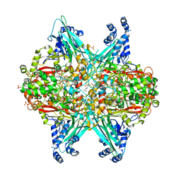
6UBZ
DownloadVisualize
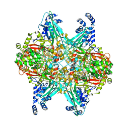
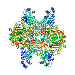
BU of 6ubz by Molmil
Crystal structure of D678A GoxA bound to glycine at pH 5.5
分子名称: GLYCINE, MAGNESIUM ION, Uncharacterized protein GoxA
著者Yukl, E.T.
登録日2019-09-13
公開日2019-10-23
最終更新日2023-10-11
実験手法X-RAY DIFFRACTION (1.83 Å)
主引用文献Kinetic and structural evidence that Asp-678 plays multiple roles in catalysis by the quinoprotein glycine oxidase.
J.Biol.Chem., 294, 2019
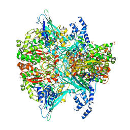
6UC1
DownloadVisualize
BU of 6uc1 by Molmil
Crystal structure of D678A GoxA soaked in glycine at pH 7.5
分子名称: GLYCINE, MAGNESIUM ION, SULFATE ION, ...
著者Yukl, E.T.
登録日2019-09-13
公開日2019-10-23
最終更新日2023-10-11
実験手法X-RAY DIFFRACTION (2.19 Å)
主引用文献Kinetic and structural evidence that Asp-678 plays multiple roles in catalysis by the quinoprotein glycine oxidase.
J.Biol.Chem., 294, 2019
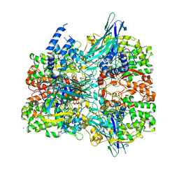
6VMF
DownloadVisualize
BU of 6vmf by Molmil
Crystal structure of the Y766F mutant of GoxA soaked with glycine
分子名称: Glycine oxidase, MAGNESIUM ION, SULFATE ION
著者Yukl, E.T.
登録日2020-01-27
公開日2020-04-08
最終更新日2023-10-11
実験手法X-RAY DIFFRACTION (2.24 Å)
主引用文献Roles of active-site residues in catalysis, substrate binding, cooperativity, and the reaction mechanism of the quinoprotein glycine oxidase.
J.Biol.Chem., 295, 2020
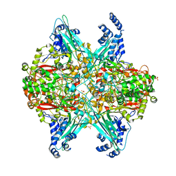
6VMW
DownloadVisualize
BU of 6vmw by Molmil
Crystal structure of the F316A mutant of GoxA soaked with glycine
分子名称: Glycine oxidase, MAGNESIUM ION, SODIUM ION
著者Yukl, E.T.
登録日2020-01-28
公開日2020-04-08
最終更新日2023-10-11
実験手法X-RAY DIFFRACTION (1.99 Å)
主引用文献Roles of active-site residues in catalysis, substrate binding, cooperativity, and the reaction mechanism of the quinoprotein glycine oxidase.
J.Biol.Chem., 295, 2020
6VMV
DownloadVisualize
BU of 6vmv by Molmil
Crystal structure of the H767A mutant of GoxA soaked with glycine
分子名称: Glycine oxidase, MAGNESIUM ION, SULFATE ION
著者Yukl, E.T.
登録日2020-01-28
公開日2020-04-08
最終更新日2023-10-11
実験手法X-RAY DIFFRACTION (2.2 Å)
主引用文献Roles of active-site residues in catalysis, substrate binding, cooperativity, and the reaction mechanism of the quinoprotein glycine oxidase.
J.Biol.Chem., 295, 2020
6VL7
DownloadVisualize
BU of 6vl7 by Molmil
Crystal structure of the H583C mutant of GoxA soaked with glycine
分子名称: DI(HYDROXYETHYL)ETHER, Glycine oxidase, MAGNESIUM ION, ...
著者Yukl, E.T.
登録日2020-01-22
公開日2020-04-08
最終更新日2023-10-11
実験手法X-RAY DIFFRACTION (2.14 Å)
主引用文献Roles of active-site residues in catalysis, substrate binding, cooperativity, and the reaction mechanism of the quinoprotein glycine oxidase.
J.Biol.Chem., 295, 2020
1AAC
DownloadVisualize
BU of 1aac by Molmil
AMICYANIN OXIDIZED, 1.31 ANGSTROMS
分子名称: AMICYANIN, COPPER (II) ION
著者Cunane, L.M, Chen, Z.-W, Durley, R.C.E, Mathews, F.S.
登録日1995-09-07
公開日1996-03-08
最終更新日2024-02-07
実験手法X-RAY DIFFRACTION (1.31 Å)
主引用文献X-ray structure of the cupredoxin amicyanin, from Paracoccus denitrificans, refined at 1.31 A resolution.
Acta Crystallogr.,Sect.D, 52, 1996
2MAD
DownloadVisualize
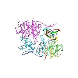
BU of 2mad by Molmil
THE ACTIVE SITE STRUCTURE OF METHYLAMINE DEHYDROGENASE: HYDRAZINES IDENTIFY C6 AS THE REACTIVE SITE OF THE TRYPTOPHAN DERIVED QUINONE COFACTOR
分子名称: METHYLAMINE DEHYDROGENASE (HEAVY SUBUNIT), METHYLAMINE DEHYDROGENASE (LIGHT SUBUNIT)
著者Huizinga, E.G, Vellieux, F.M.D, Hol, W.G.J.
登録日1992-05-20
公開日1994-01-31
最終更新日2024-06-05
実験手法X-RAY DIFFRACTION (2.25 Å)
主引用文献Active site structure of methylamine dehydrogenase: hydrazines identify C6 as the reactive site of the tryptophan-derived quinone cofactor.
Biochemistry, 31, 1992
1MAE
DownloadVisualize
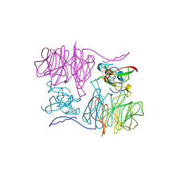
BU of 1mae by Molmil
The Active Site Structure of Methylamine Dehydrogenase: Hydrazines Identify C6 as the Reactive Site of the Tryptophan Derived Quinone Cofactor
分子名称: METHYLAMINE DEHYDROGENASE (HEAVY SUBUNIT), METHYLAMINE DEHYDROGENASE (LIGHT SUBUNIT), NITROGEN MOLECULE
著者Huizinga, E.G, Vellieux, F.M.D, Hol, W.G.J.
登録日1992-05-20
公開日1994-01-31
最終更新日2024-06-05
実験手法X-RAY DIFFRACTION (2.8 Å)
主引用文献Active site structure of methylamine dehydrogenase: hydrazines identify C6 as the reactive site of the tryptophan-derived quinone cofactor.
Biochemistry, 31, 1992
1MAF
DownloadVisualize
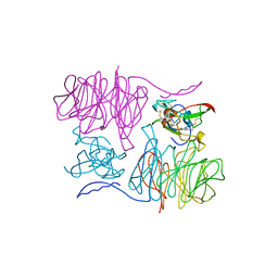
BU of 1maf by Molmil
The Active Site Structure of Methylamine Dehydrogenase: Hydrazines Identify C6 as the Reactive Site of the Tryptophan Derived Quinone Cofactor
分子名称: METHYLAMINE DEHYDROGENASE (HEAVY SUBUNIT), METHYLAMINE DEHYDROGENASE (LIGHT SUBUNIT), NITROGEN MOLECULE
著者Huizinga, E.G, Vellieux, F.M.D, Hol, W.G.J.
登録日1992-05-20
公開日1994-01-31
最終更新日2024-06-05
実験手法X-RAY DIFFRACTION (2.6 Å)
主引用文献Active site structure of methylamine dehydrogenase: hydrazines identify C6 as the reactive site of the tryptophan-derived quinone cofactor.
Biochemistry, 31, 1992
<1234


238582

件を2025-07-09に公開中

PDB statisticsPDBj update infoContact PDBjnumon