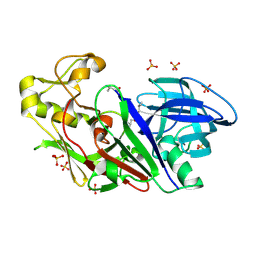
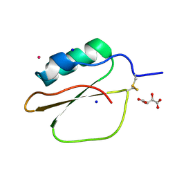
2DDB
 
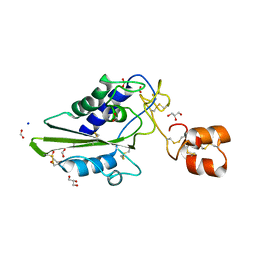 | | Crystal structure of pseudecin from Pseudechis porphyriacus | | 分子名称: | FORMIC ACID, GLYCEROL, Pseudecin, ... | | 著者 | Suzuki, N, Yamazaki, Y, Fujimoto, Z, Morita, T, Mizuno, H. | | 登録日 | 2006-01-25 | | 公開日 | 2007-01-30 | | 最終更新日 | 2024-11-20 | | 実験手法 | X-RAY DIFFRACTION (1.9 Å) | | 主引用文献 | Structures of pseudechetoxin and pseudecin, two snake-venom cysteine-rich secretory proteins that target cyclic nucleotide-gated ion channels: implications for movement of the C-terminal cysteine-rich domain
Acta Crystallogr.,Sect.D, 64, 2008
|
|
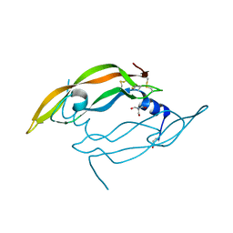
2DDA
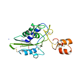 | | Crystal structure of pseudechetoxin from Pseudechis australis | | 分子名称: | FORMIC ACID, GLYCEROL, Pseudechetoxin, ... | | 著者 | Suzuki, N, Yamazaki, Y, Fujimoto, Z, Morita, T, Mizuno, H. | | 登録日 | 2006-01-25 | | 公開日 | 2007-01-30 | | 最終更新日 | 2024-10-30 | | 実験手法 | X-RAY DIFFRACTION (2.25 Å) | | 主引用文献 | Structures of pseudechetoxin and pseudecin, two snake-venom cysteine-rich secretory proteins that target cyclic nucleotide-gated ion channels: implications for movement of the C-terminal cysteine-rich domain
Acta Crystallogr.,Sect.D, 64, 2008
|
|
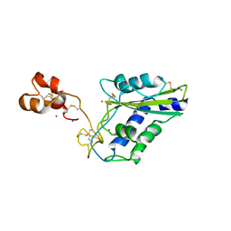
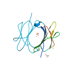
1WDU
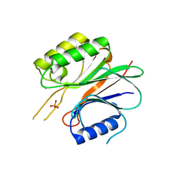 | | Endonuclease domain of TRAS1, a telomere-specific non-LTR retrotransposon | | 分子名称: | CHLORIDE ION, PHOSPHATE ION, TRAS1 ORF2p | | 著者 | Maita, N, Anzai, T, Aoyagi, H, Mizuno, H, Fujiwara, H. | | 登録日 | 2004-05-17 | | 公開日 | 2004-08-10 | | 最終更新日 | 2024-05-29 | | 実験手法 | X-RAY DIFFRACTION (2.4 Å) | | 主引用文献 | Crystal structure of the endonuclease domain encoded by the telomere-specific long interspersed nuclear element, TRAS1
J.Biol.Chem., 279, 2004
|
|
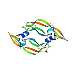
2EPF
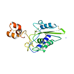 | | Crystal Structure of Zinc-Bound Pseudecin From Pseudechis Porphyriacus | | 分子名称: | Pseudecin, SODIUM ION, ZINC ION | | 著者 | Suzuki, N, Yamazaki, Y, Fujimoto, Z, Morita, T, Mizuno, H. | | 登録日 | 2007-03-29 | | 公開日 | 2008-03-11 | | 最終更新日 | 2024-11-20 | | 実験手法 | X-RAY DIFFRACTION (2.3 Å) | | 主引用文献 | Structures of pseudechetoxin and pseudecin, two snake-venom cysteine-rich secretory proteins that target cyclic nucleotide-gated ion channels: implications for movement of the C-terminal cysteine-rich domain
Acta Crystallogr.,Sect.D, 64, 2008
|
|
2GW3
 | | Crystal structure of stony coral fluorescent protein Kaede, green form | | 分子名称: | Kaede, NICKEL (II) ION | | 著者 | Hayashi, I, Mizuno, H, Miyawaki, A, Ikura, M. | | 登録日 | 2006-05-03 | | 公開日 | 2007-05-08 | | 最終更新日 | 2024-10-30 | | 実験手法 | X-RAY DIFFRACTION (1.4 Å) | | 主引用文献 | Crystallographic evidence for water-assisted photo-induced peptide cleavage in the stony coral fluorescent protein Kaede.
J.Mol.Biol., 372, 2007
|
|
2GW4
 | | Crystal structure of stony coral fluorescent protein Kaede, red form | | 分子名称: | Kaede, NICKEL (II) ION | | 著者 | Hayashi, I, Mizuno, H, Miyawako, A, Ikura, M. | | 登録日 | 2006-05-03 | | 公開日 | 2007-05-08 | | 最終更新日 | 2023-11-15 | | 実験手法 | X-RAY DIFFRACTION (1.6 Å) | | 主引用文献 | Crystallographic evidence for water-assisted photo-induced peptide cleavage in the stony coral fluorescent protein Kaede.
J.Mol.Biol., 372, 2007
|
|
2ZMU
 | | Crystal Structure of Monomeric Kusabira-Orange (MKO), Orange-Emitting GFP-like Protein, at pH 9.1 | | 分子名称: | Fluorescent protein | | 著者 | Kikuchi, A, Fukumura, E, Karasawa, S, Mizuno, H, Miyawaki, A, Shiro, Y, RIKEN Structural Genomics/Proteomics Initiative (RSGI) | | 登録日 | 2008-04-21 | | 公開日 | 2008-10-21 | | 最終更新日 | 2024-10-16 | | 実験手法 | X-RAY DIFFRACTION (1.65 Å) | | 主引用文献 | Structural Characterization of a Thiazoline-Containing Chromophore in an Orange Fluorescent Protein, Monomeric Kusabira Orange
Biochemistry, 47, 2008
|
|
2ZMW
 | | Crystal Structure of Monomeric Kusabira-Orange (MKO), Orange-Emitting GFP-like Protein, at pH 6.0 | | 分子名称: | Fluorescent protein | | 著者 | Kikuchi, A, Fukumura, E, Karasawa, S, Mizuno, H, Miyawaki, A, Shiro, Y, RIKEN Structural Genomics/Proteomics Initiative (RSGI) | | 登録日 | 2008-04-21 | | 公開日 | 2008-10-21 | | 最終更新日 | 2024-10-16 | | 実験手法 | X-RAY DIFFRACTION (2 Å) | | 主引用文献 | Structural Characterization of a Thiazoline-Containing Chromophore in an Orange Fluorescent Protein, Monomeric Kusabira Orange
Biochemistry, 47, 2008
|
|
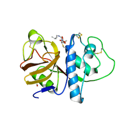
1V6V
 | | Crystal Structure Of Xylanase From Streptomyces Olivaceoviridis E-86 Complexed With 3(2)-alpha-L-arabinofuranosyl-xylotriose | | 分子名称: | ENDO-1,4-BETA-D-XYLANASE, alpha-L-arabinofuranose-(1-3)-[beta-D-xylopyranose-(1-4)]beta-D-xylopyranose-(1-4)-beta-D-xylopyranose, beta-D-xylopyranose, ... | | 著者 | Fujimoto, Z, Kaneko, S, Kuno, A, Kobayashi, H, Kusakabe, I, Mizuno, H. | | 登録日 | 2003-12-04 | | 公開日 | 2004-04-27 | | 最終更新日 | 2024-10-23 | | 実験手法 | X-RAY DIFFRACTION (2.1 Å) | | 主引用文献 | Crystal structures of decorated xylooligosaccharides bound to a family 10 xylanase from Streptomyces olivaceoviridis E-86
J.Biol.Chem., 279, 2004
|
|
1V6X
 | | Crystal Structure Of Xylanase From Streptomyces Olivaceoviridis E-86 Complexed With 3(3)-4-O-methyl-alpha-D-glucuronosyl-xylotriose | | 分子名称: | 4-O-methyl-alpha-D-glucopyranuronic acid-(1-2)-beta-D-xylopyranose-(1-4)-beta-D-xylopyranose-(1-4)-beta-D-xylopyranose, ENDO-1,4-BETA-D-XYLANASE, beta-D-xylopyranose, ... | | 著者 | Fujimoto, Z, Kaneko, S, Kuno, A, Kobayashi, H, Kusakabe, I, Mizuno, H. | | 登録日 | 2003-12-04 | | 公開日 | 2004-04-27 | | 最終更新日 | 2024-11-13 | | 実験手法 | X-RAY DIFFRACTION (2.1 Å) | | 主引用文献 | Crystal structures of decorated xylooligosaccharides bound to a family 10 xylanase from Streptomyces olivaceoviridis E-86
J.Biol.Chem., 279, 2004
|
|
1V6U
 | | Crystal Structure Of Xylanase From Streptomyces Olivaceoviridis E-86 Complexed With 2(2)-alpha-L-arabinofuranosyl-xylobiose | | 分子名称: | alpha-D-xylopyranose, alpha-L-arabinofuranose-(1-3)-beta-D-xylopyranose-(1-4)-beta-D-xylopyranose, beta-D-xylopyranose, ... | | 著者 | Fujimoto, Z, Kaneko, S, Kuno, A, Kobayashi, H, Kusakabe, I, Mizuno, H. | | 登録日 | 2003-12-04 | | 公開日 | 2004-04-27 | | 最終更新日 | 2024-11-20 | | 実験手法 | X-RAY DIFFRACTION (2.1 Å) | | 主引用文献 | Crystal structures of decorated xylooligosaccharides bound to a family 10 xylanase from Streptomyces olivaceoviridis E-86
J.Biol.Chem., 279, 2004
|
|
1V6W
 | | Crystal Structure Of Xylanase From Streptomyces Olivaceoviridis E-86 Complexed With 2(2)-4-O-methyl-alpha-D-glucuronosyl-xylobiose | | 分子名称: | 4-O-methyl-alpha-D-glucopyranuronic acid-(1-2)-beta-D-xylopyranose-(1-4)-beta-D-xylopyranose, ENDO-1,4-BETA-D-XYLANASE, beta-D-xylopyranose, ... | | 著者 | Fujimoto, Z, Kaneko, S, Kuno, A, Kobayashi, H, Kusakabe, I, Mizuno, H. | | 登録日 | 2003-12-04 | | 公開日 | 2004-04-27 | | 最終更新日 | 2024-11-20 | | 実験手法 | X-RAY DIFFRACTION (2 Å) | | 主引用文献 | Crystal structures of decorated xylooligosaccharides bound to a family 10 xylanase from Streptomyces olivaceoviridis E-86
J.Biol.Chem., 279, 2004
|
|
1WKR
 | | Crystal structure of aspartic proteinase from Irpex lacteus | | 分子名称: | Polyporopepsin, SULFATE ION, pepstatin | | 著者 | Fujimoto, Z, Fujii, Y, Kaneko, S, Kobayashi, H, Mizuno, H. | | 登録日 | 2004-06-02 | | 公開日 | 2004-09-07 | | 最終更新日 | 2024-10-16 | | 実験手法 | X-RAY DIFFRACTION (1.3 Å) | | 主引用文献 | Crystal Structure of Aspartic Proteinase from Irpex lacteus in Complex with Inhibitor Pepstatin
J.Mol.Biol., 341, 2004
|
|
1WQ8
 | | Crystal structure of Vammin, a VEGF-F from a snake venom | | 分子名称: | 2-AMINO-2-HYDROXYMETHYL-PROPANE-1,3-DIOL, Vascular endothelial growth factor toxin | | 著者 | Suto, K, Yamazaki, Y, Morita, T, Mizuno, H. | | 登録日 | 2004-09-23 | | 公開日 | 2004-12-07 | | 最終更新日 | 2024-10-23 | | 実験手法 | X-RAY DIFFRACTION (1.9 Å) | | 主引用文献 | Crystal structures of novel vascular endothelial growth factors (VEGF) from snake venoms: insight into selective VEGF binding to kinase insert domain-containing receptor but not to fms-like tyrosine kinase-1.
J.Biol.Chem., 280, 2005
|
|
1WVR
 | | Crystal Structure of a CRISP family Ca-channel blocker derived from snake venom | | 分子名称: | CADMIUM ION, Triflin | | 著者 | Shikamoto, Y, Suto, K, Yamazaki, Y, Morita, T, Mizuno, H. | | 登録日 | 2004-12-24 | | 公開日 | 2005-07-05 | | 最終更新日 | 2024-10-23 | | 実験手法 | X-RAY DIFFRACTION (2.4 Å) | | 主引用文献 | Crystal structure of a CRISP family Ca2+ -channel blocker derived from snake venom.
J.Mol.Biol., 350, 2005
|
|
1WQ9
 | | Crystal structure of VR-1, a VEGF-F from a snake venom | | 分子名称: | Vascular endothelial growth factor | | 著者 | Suto, K, Yamazaki, Y, Morita, T, Mizuno, H. | | 登録日 | 2004-09-24 | | 公開日 | 2004-12-07 | | 最終更新日 | 2024-10-23 | | 実験手法 | X-RAY DIFFRACTION (2 Å) | | 主引用文献 | Crystal structures of novel vascular endothelial growth factors (VEGF) from snake venoms: insight into selective VEGF binding to kinase insert domain-containing receptor but not to fms-like tyrosine kinase-1.
J.Biol.Chem., 280, 2005
|
|
1X2W
 | | Crystal Structure of Apo-Habu IX-bp at pH 4.6 | | 分子名称: | CHLORIDE ION, Coagulation factor IX/X-binding protein A chain, Coagulation factor IX/factor X-binding protein B chain, ... | | 著者 | Suzuki, N, Fujimoto, Z, Morita, T, Fukamizu, A, Mizuno, H. | | 登録日 | 2005-04-26 | | 公開日 | 2005-10-04 | | 最終更新日 | 2024-10-16 | | 実験手法 | X-RAY DIFFRACTION (2.29 Å) | | 主引用文献 | pH-Dependent Structural Changes at Ca(2+)-binding sites of Coagulation Factor IX-binding Protein
J.Mol.Biol., 353, 2005
|
|
1WQZ
 | | Complicated water orientations in the minor groove of B-DNA decamer D(CCATTAATGG)2 observed by neutron diffraction measurements | | 分子名称: | 5'-D(*CP*CP*AP*TP*TP*AP*AP*TP*GP*G)-3' | | 著者 | Arai, S, Chatake, T, Ohhara, T, Kurihara, K, Tanaka, I, Suzuki, N, Fujimoto, Z, Mizuno, H, Niimura, N. | | 登録日 | 2004-10-07 | | 公開日 | 2005-06-21 | | 最終更新日 | 2023-10-25 | | 実験手法 | NEUTRON DIFFRACTION (3 Å) | | 主引用文献 | Complicated water orientations in the minor groove of the B-DNA decamer d(CCATTAATGG)2 observed by neutron diffraction measurements
Nucleic Acids Res., 33, 2005
|
|
1WQY
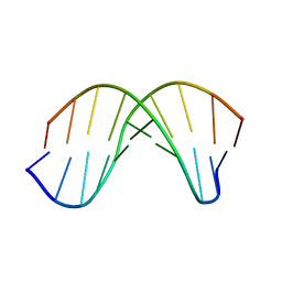 | | X-RAY structural analysis of B-DNA decamer D(CCATTAATGG)2 crystal grown in D2O solution | | 分子名称: | 5'-D(*CP*CP*AP*TP*TP*AP*AP*TP*GP*G)-3' | | 著者 | Arai, S, Chatake, T, Ohhara, T, Kurihara, K, Tanaka, I, Suzuki, N, Fujimoto, Z, Mizuno, H, Niimura, N. | | 登録日 | 2004-10-07 | | 公開日 | 2005-06-21 | | 最終更新日 | 2023-10-25 | | 実験手法 | X-RAY DIFFRACTION (2 Å) | | 主引用文献 | Complicated water orientations in the minor groove of the B-DNA decamer d(CCATTAATGG)2 observed by neutron diffraction measurements
Nucleic Acids Res., 33, 2005
|
|
1X2T
 | | Crystal Structure of Habu IX-bp at pH 6.5 | | 分子名称: | CALCIUM ION, Coagulation factor IX/X-binding protein A chain, Coagulation factor IX/factor X-binding protein B chain, ... | | 著者 | Suzuki, N, Fujimoto, Z, Morita, T, Fukamizu, A, Mizuno, H. | | 登録日 | 2005-04-26 | | 公開日 | 2005-10-04 | | 最終更新日 | 2024-10-16 | | 実験手法 | X-RAY DIFFRACTION (1.72 Å) | | 主引用文献 | pH-Dependent Structural Changes at Ca(2+)-binding sites of Coagulation Factor IX-binding Protein
J.Mol.Biol., 353, 2005
|
|
1VBW
 | | Crystal Structure of Bitter Gourd Trypsin Inhibitor | | 分子名称: | L(+)-TARTARIC ACID, POTASSIUM ION, SODIUM ION, ... | | 著者 | Suto, K, Furuichi, M, Nishimoto, E, Meno, K, Horii, K, Mizuno, H. | | 登録日 | 2004-03-03 | | 公開日 | 2005-03-22 | | 最終更新日 | 2024-10-30 | | 実験手法 | X-RAY DIFFRACTION (0.93 Å) | | 主引用文献 | Crystal Structure of Bitter Gourd Trypsin Inhibitor
to be published
|
|
6M1B
 | | A new V27M variant of beta 2 microglobulin induced amyloidosis in a patient with long-term hemodialysis | | 分子名称: | Beta-2-microglobulin, CALCIUM ION, GLYCEROL, ... | | 著者 | So, M, Nakahara, S, Nakaniwa, T, Tanaka, H, Kurisu, G, Goto, Y. | | 登録日 | 2020-02-25 | | 公開日 | 2021-01-06 | | 最終更新日 | 2024-10-16 | | 実験手法 | X-RAY DIFFRACTION (1.88 Å) | | 主引用文献 | Dialysis-related amyloidosis associated with a novel beta 2 -microglobulin variant.
Amyloid, 28, 2021
|
|
1PPP
 | |
1TRA
 | | RESTRAINED REFINEMENT OF THE MONOCLINIC FORM OF YEAST PHENYLALANINE TRANSFER RNA. TEMPERATURE FACTORS AND DYNAMICS, COORDINATED WATERS, AND BASE-PAIR PROPELLER TWIST ANGLES | | 分子名称: | MAGNESIUM ION, TRNAPHE | | 著者 | Westhof, E, Sundaralingam, M. | | 登録日 | 1986-05-16 | | 公開日 | 1986-07-14 | | 最終更新日 | 2024-02-14 | | 実験手法 | X-RAY DIFFRACTION (3 Å) | | 主引用文献 | Restrained refinement of the monoclinic form of yeast phenylalanine transfer RNA. Temperature factors and dynamics, coordinated waters, and base-pair propeller twist angles.
Biochemistry, 25, 1986
|
|
3BOY
 | | Crystal structure of the HutP antitermination complex bound to the HUT mRNA | | 分子名称: | 5'-R(*UP*UP*UP*AP*GP*UP*UP*UP*UP*UP*AP*GP*UP*UP*UP*UP*UP*AP*GP*UP*UP*U)-3', HISTIDINE, Hut operon positive regulatory protein, ... | | 著者 | Kumarevel, T.S, Balasundaresan, D, Jeyakanthan, J, Shinkai, A, Yokoyama, S, Kumar, P.K.R, RIKEN Structural Genomics/Proteomics Initiative (RSGI) | | 登録日 | 2007-12-18 | | 公開日 | 2008-01-15 | | 最終更新日 | 2023-11-01 | | 実験手法 | X-RAY DIFFRACTION (1.7 Å) | | 主引用文献 | Crystal Structure of HutP complexed with the 55-mer RNA
To be Published
|
|


