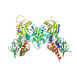
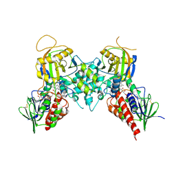
8W22
 
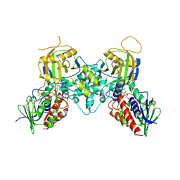
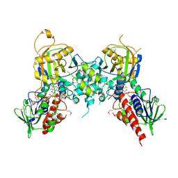
 | | Umb1 umbrella toxin particle (local refinement of UmbB1 bound ALF of UmbC1 and UmbA1) | | 分子名称: | Intein C-terminal splicing domain-containing protein, Secreted esterase, Secreted protein | | 著者 | Park, Y.J, Zhao, Q, Seattle Structural Genomics Center for Infectious Disease (SSGCID), DiMaio, F, Mougous, J.D, Veesler, D. | | 登録日 | 2024-02-19 | | 公開日 | 2024-04-17 | | 最終更新日 | 2024-05-15 | | 実験手法 | ELECTRON MICROSCOPY (4 Å) | | 主引用文献 | Streptomyces umbrella toxin particles block hyphal growth of competing species.
Nature, 629, 2024
|
|
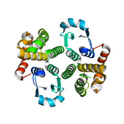
8W20
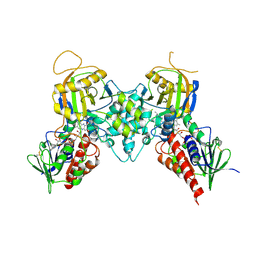
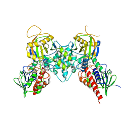
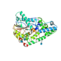
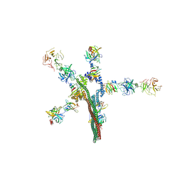 | | Umb1 umbrella toxin particle | | 分子名称: | Intein C-terminal splicing domain-containing protein, Secreted esterase, Secreted protein | | 著者 | Park, Y.J, Zhao, Q, Seattle Structural Genomics Center for Infectious Disease (SSGCID), DiMaio, F, Mougous, J.D, Veesler, D. | | 登録日 | 2024-02-19 | | 公開日 | 2024-04-17 | | 最終更新日 | 2024-05-15 | | 実験手法 | ELECTRON MICROSCOPY (4.3 Å) | | 主引用文献 | Streptomyces umbrella toxin particles block hyphal growth of competing species.
Nature, 629, 2024
|
|
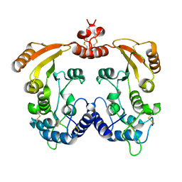
3CN8
 | |
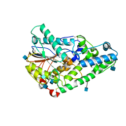
5WTD
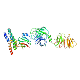
 | | Structure of human serum transferrin bound ruthenium at N-lobe | | 分子名称: | FE (III) ION, MALONATE ION, RUTHENIUM ION, ... | | 著者 | Sun, H, Wang, M, Lai, T.P, Zhang, H, Hao, Q. | | 登録日 | 2016-12-11 | | 公開日 | 2017-12-20 | | 最終更新日 | 2023-11-08 | | 実験手法 | X-RAY DIFFRACTION (2.501 Å) | | 主引用文献 | Binding of ruthenium and osmium at non‐iron sites of transferrin accounts for their iron-independent cellular uptake.
J.Inorg.Biochem., 234, 2022
|
|
2B5H
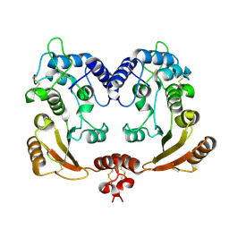
 | | 1.5 A Resolution Crystal Structure of Recombinant R. Norvegicus Cysteine Dioxygenase | | 分子名称: | Cysteine dioxygenase type I, FE (III) ION | | 著者 | Simmons, C.R, Liu, Q, Huang, Q, Hao, Q, Begley, T.P, Karplus, P.A, Stipanuk, M.H. | | 登録日 | 2005-09-28 | | 公開日 | 2006-04-11 | | 最終更新日 | 2024-02-14 | | 実験手法 | X-RAY DIFFRACTION (1.5 Å) | | 主引用文献 | Crystal Structure of Mammalian Cysteine Dioxygenase: A NOVEL MONONUCLEAR IRON CENTER FOR CYSTEINE THIOL OXIDATION.
J.Biol.Chem., 281, 2006
|
|
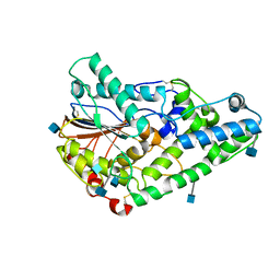
1QWO
 | | Crystal structure of a phosphorylated phytase from Aspergillus fumigatus, revealing the structural basis for its heat resilience and catalytic pathway | | 分子名称: | 2-acetamido-2-deoxy-beta-D-glucopyranose, phytase | | 著者 | Xiang, T, Liu, Q, Deacon, A.M, Koshy, M, Kriksunov, I.A, Lei, X.G, Hao, Q, Thiel, D.J. | | 登録日 | 2003-09-03 | | 公開日 | 2004-06-01 | | 最終更新日 | 2020-07-29 | | 実験手法 | X-RAY DIFFRACTION (1.5 Å) | | 主引用文献 | Crystal Structure of a Heat-resilient Phytase from Aspergillus fumigatus, Carrying a Phosphorylated Histidine
J.Mol.Biol., 339, 2004
|
|
1TW9
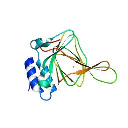
 | | Glutathione Transferase-2, apo form, from the nematode Heligmosomoides polygyrus | | 分子名称: | glutathione S-transferase 2 | | 著者 | Schuller, D.J, Liu, Q, Kriksunov, I.A, Campbell, A.M, Barrett, J, Brophy, P.M, Hao, Q. | | 登録日 | 2004-06-30 | | 公開日 | 2004-08-03 | | 最終更新日 | 2023-08-23 | | 実験手法 | X-RAY DIFFRACTION (1.71 Å) | | 主引用文献 | Crystal structure of a new class of glutathione transferase from the model human hookworm nematode Heligmosomoides polygyrus.
Proteins, 61, 2005
|
|
3BI5
 | |
3BI4
 | |
3BI2
 | |
3BNM
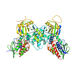
 | | Crystal structure of polyamine oxidase FMS1 from Saccharomyces cerevisiae in complex with bis-(3R,3'R)-methylated spermine | | 分子名称: | (3R,3'R)-N~1~,N~1~'-butane-1,4-diyldibutane-1,3-diamine, FLAVIN-ADENINE DINUCLEOTIDE, Polyamine oxidase FMS1 | | 著者 | Huang, Q, Hao, Q. | | 登録日 | 2007-12-14 | | 公開日 | 2008-01-29 | | 最終更新日 | 2011-07-13 | | 実験手法 | X-RAY DIFFRACTION (2.2 Å) | | 主引用文献 | Structural basis of the substrate stereospecificity of FMS1.
To Be Published
|
|
3BNU
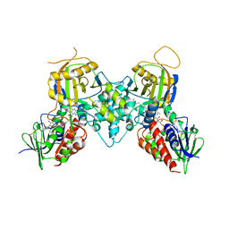
 | | Crystal structure of polyamine oxidase FMS1 from Saccharomyces cerevisiae in complex with bis-(3S,3'S)-methylated spermine | | 分子名称: | (3S,3'S)-N~1~,N~1~'-butane-1,4-diyldibutane-1,3-diamine, FLAVIN-ADENINE DINUCLEOTIDE, Polyamine oxidase FMS1 | | 著者 | Huang, Q, Hao, Q. | | 登録日 | 2007-12-14 | | 公開日 | 2008-01-29 | | 最終更新日 | 2011-07-13 | | 実験手法 | X-RAY DIFFRACTION (2.2 Å) | | 主引用文献 | Structural basis of the substrate stereospecificity of FMS1.
To Be Published
|
|
1R12
 | | Native Aplysia ADP ribosyl cyclase | | 分子名称: | ADP-ribosyl cyclase | | 著者 | Love, M.L, Szebenyi, D.M.E, Kriksunov, I.A, Thiel, D.J, Munshi, C, Graeff, R, Lee, H.C, Hao, Q. | | 登録日 | 2003-09-23 | | 公開日 | 2004-03-09 | | 最終更新日 | 2011-07-13 | | 実験手法 | X-RAY DIFFRACTION (1.7 Å) | | 主引用文献 | ADP-ribosyl cyclase; crystal structures reveal a covalent intermediate.
Structure, 12, 2004
|
|
1R0S
 | | Crystal structure of ADP-ribosyl cyclase Glu179Ala mutant | | 分子名称: | ADP-ribosyl cyclase | | 著者 | Love, M.L, Szebenyi, D.M.E, Kriksunov, I.A, Thiel, D.J, Munshi, C, Graeff, R, Lee, H.C, Hao, Q. | | 登録日 | 2003-09-22 | | 公開日 | 2004-03-09 | | 最終更新日 | 2021-10-27 | | 実験手法 | X-RAY DIFFRACTION (2 Å) | | 主引用文献 | ADP-ribosyl cyclase; crystal structures reveal a covalent intermediate.
Structure, 12, 2004
|
|
3CNP
 | |
3CND
 | |
3CNT
 | |
1SKB
 | | Crystallographic snapshots of Aspergillus fumigatus phytase revealing its enzymatic dynamics | | 分子名称: | 2-acetamido-2-deoxy-beta-D-glucopyranose, 3-phytase A | | 著者 | Liu, Q, Huang, Q, Lei, X.G, Hao, Q. | | 登録日 | 2004-03-04 | | 公開日 | 2004-09-28 | | 最終更新日 | 2020-07-29 | | 実験手法 | X-RAY DIFFRACTION (1.58 Å) | | 主引用文献 | Crystallographic Snapshots of Aspergillus fumigatus Phytase, Revealing Its Enzymatic Dynamics
Structure, 12, 2004
|
|
1REO
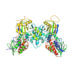
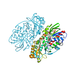 | | L-amino acid oxidase from Agkistrodon halys pallas | | 分子名称: | 2-acetamido-2-deoxy-beta-D-glucopyranose, AHPLAAO, CITRIC ACID, ... | | 著者 | Zhang, H, Teng, M, Niu, L, Wang, Y, Wang, Y, Liu, Q, Huang, Q, Hao, Q, Dong, Y, Liu, P. | | 登録日 | 2003-11-07 | | 公開日 | 2004-05-04 | | 最終更新日 | 2023-10-25 | | 実験手法 | X-RAY DIFFRACTION (2.31 Å) | | 主引用文献 | Purification, partial characterization, crystallization and structural determination of AHP-LAAO, a novel L-amino-acid oxidase with cell apoptosis-inducing activity from Agkistrodon halys pallas venom.
Acta Crystallogr.,Sect.D, 60, 2004
|
|
1SKA
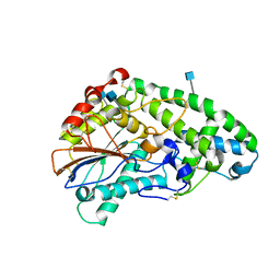 | | Crystallographic snapshots of Aspergillus fumigatus phytase revealing its enzymatic dynamics | | 分子名称: | 2-acetamido-2-deoxy-beta-D-glucopyranose, 3-phytase A | | 著者 | Liu, Q, Huang, Q, Lei, X.G, Hao, Q. | | 登録日 | 2004-03-04 | | 公開日 | 2004-09-28 | | 最終更新日 | 2020-07-29 | | 実験手法 | X-RAY DIFFRACTION (1.69 Å) | | 主引用文献 | Crystallographic Snapshots of Aspergillus fumigatus Phytase, Revealing Its Enzymatic Dynamics
Structure, 12, 2004
|
|
1SK9
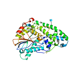 | | Crystallographic snapshots of Aspergillus fumigatus phytase revealing its enzymatic dynamics | | 分子名称: | 2-acetamido-2-deoxy-beta-D-glucopyranose, 3-phytase A, PHOSPHATE ION | | 著者 | Liu, Q, Huang, Q, Lei, X.G, Hao, Q. | | 登録日 | 2004-03-04 | | 公開日 | 2004-09-28 | | 最終更新日 | 2020-07-29 | | 実験手法 | X-RAY DIFFRACTION (1.64 Å) | | 主引用文献 | Crystallographic Snapshots of Aspergillus fumigatus Phytase, Revealing Its Enzymatic Dynamics
Structure, 12, 2004
|
|
1RSG
 | |
1VB0
 | | Atomic resolution structure of atratoxin-b, one short-chain neurotoxin from Naja atra | | 分子名称: | 2-AMINO-2-HYDROXYMETHYL-PROPANE-1,3-DIOL, Cobrotoxin b, SULFATE ION | | 著者 | Lou, X, Liu, Q, Teng, M, Niu, L, Huang, Q, Hao, Q. | | 登録日 | 2004-02-20 | | 公開日 | 2004-12-21 | | 最終更新日 | 2023-10-25 | | 実験手法 | X-RAY DIFFRACTION (0.92 Å) | | 主引用文献 | The atomic resolution crystal structure of atratoxin determined by single wavelength anomalous diffraction phasing
J.Biol.Chem., 279, 2004
|
|
1V6P
 | | Crystal structure of Cobrotoxin | | 分子名称: | CHLORIDE ION, COPPER (II) ION, Cobrotoxin, ... | | 著者 | Lou, X, Tu, X, Wang, J, Teng, M, Niu, L, Liu, Q, Huang, Q, Hao, Q. | | 登録日 | 2003-12-03 | | 公開日 | 2004-12-21 | | 最終更新日 | 2023-12-27 | | 実験手法 | X-RAY DIFFRACTION (0.87 Å) | | 主引用文献 | The atomic resolution crystal structure of atratoxin determined by single wavelength anomalous diffraction phasing
J.Biol.Chem., 279, 2004
|
|
1SK8
 | | Crystallographic snapshots of Aspergillus fumigatus phytase revealing its enzymatic dynamics | | 分子名称: | 2-acetamido-2-deoxy-beta-D-glucopyranose, 3-phytase A, PHOSPHATE ION | | 著者 | Liu, Q, Huang, Q.Q, Lei, X.G, Hao, Q. | | 登録日 | 2004-03-04 | | 公開日 | 2005-02-08 | | 最終更新日 | 2020-07-29 | | 実験手法 | X-RAY DIFFRACTION (1.65 Å) | | 主引用文献 | Crystallographic Snapshots of Aspergillus fumigatus Phytase, Revealing Its Enzymatic Dynamics
Structure, 12, 2004
|
|


