1MCI
 
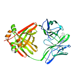 | | PRINCIPLES AND PITFALLS IN DESIGNING SITE DIRECTED PEPTIDE LIGANDS | | 分子名称: | IMMUNOGLOBULIN LAMBDA DIMER MCG (LIGHT CHAIN), PEPTIDE N-ACETYL-D-PHE-L-HIS-D-PRO-OH | | 著者 | Edmundson, A.B, Harris, D.L, Fan, Z.-C, Guddat, L.W. | | 登録日 | 1993-02-25 | | 公開日 | 1994-01-31 | | 最終更新日 | 2017-11-29 | | 実験手法 | X-RAY DIFFRACTION (2.7 Å) | | 主引用文献 | Principles and pitfalls in designing site-directed peptide ligands.
Proteins, 16, 1993
|
|
1MCS
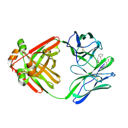 | | PRINCIPLES AND PITFALLS IN DESIGNING SITE DIRECTED PEPTIDE LIGANDS | | 分子名称: | IMMUNOGLOBULIN LAMBDA DIMER MCG (LIGHT CHAIN), PEPTIDE N-ACETYL-L-GLN-D-PHE-L-HIS-D-PRO-OH | | 著者 | Edmundson, A.B, Harris, D.L, Fan, Z.-C, Guddat, L.W. | | 登録日 | 1993-02-25 | | 公開日 | 1994-01-31 | | 最終更新日 | 2017-11-29 | | 実験手法 | X-RAY DIFFRACTION (2.7 Å) | | 主引用文献 | Principles and pitfalls in designing site-directed peptide ligands.
Proteins, 16, 1993
|
|
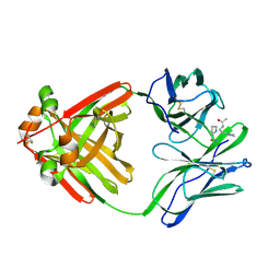
1MCE
 | | PRINCIPLES AND PITFALLS IN DESIGNING SITE DIRECTED PEPTIDE LIGANDS | | 分子名称: | Immunoglobulin lambda-1 light chain, PEPTIDE N-ACETYL-L-GLN-D-PHE-L-HIS-D-PRO-B-ALA-OH | | 著者 | Edmundson, A.B, Harris, D.L, Fan, Z.-C, Guddat, L.W. | | 登録日 | 1993-02-25 | | 公開日 | 1994-01-31 | | 最終更新日 | 2022-04-20 | | 実験手法 | X-RAY DIFFRACTION (2.7 Å) | | 主引用文献 | Principles and pitfalls in designing site-directed peptide ligands.
Proteins, 16, 1993
|
|
1MCN
 | | PRINCIPLES AND PITFALLS IN DESIGNING SITE DIRECTED PEPTIDE LIGANDS | | 分子名称: | IMMUNOGLOBULIN LAMBDA DIMER MCG (LIGHT CHAIN), PEPTIDE N-ACETYL-D-HIS-L-PRO-NH2 | | 著者 | Edmundson, A.B, Harris, D.L, Fan, Z.-C, Guddat, L.W. | | 登録日 | 1993-02-25 | | 公開日 | 1994-01-31 | | 最終更新日 | 2017-11-29 | | 実験手法 | X-RAY DIFFRACTION (2.7 Å) | | 主引用文献 | Principles and pitfalls in designing site-directed peptide ligands.
Proteins, 16, 1993
|
|


