Loading
PDBj
メニューPDBj@FacebookPDBj@TwitterPDBj@YouTubewwPDB FoundationwwPDB
RCSB PDBPDBeBMRBAdv. SearchSearch help
Search by PDB author
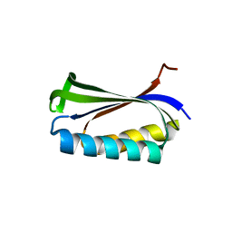
1CQM
DownloadVisualize
BU of 1cqm by Molmil
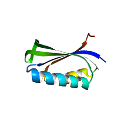
PROTEIN AGGREGATION AND ALZHEIMER'S DISEASE: CRYSTALLOGRAPHIC ANALYSIS OF THE PHENOMENON. ENGINEERED VERSION OF THE RIBOSOMAL PROTEIN S6 USED AS A STABLE SCAFFOLD TO STUDY OLIGOMERIZATION.
分子名称: RIBOSOMAL PROTEIN S6
著者Kristensen, O, Otzen, D.E, Oliveberg, M.
登録日1999-08-08
公開日2000-09-08
最終更新日2024-02-07
実験手法X-RAY DIFFRACTION (1.65 Å)
主引用文献Designed protein tetramer zipped together with a hydrophobic Alzheimer homology: a structural clue to amyloid assembly.
Proc.Natl.Acad.Sci.USA, 97, 2000
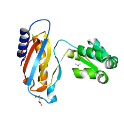
4LQ4
DownloadVisualize
BU of 4lq4 by Molmil
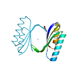
crystal structure of mutant ribosomal protein L1 from Methanococcus jannaschii with deletion of 8 residues from C-terminus
分子名称: 50S ribosomal protein L1, CHLORIDE ION, ISOPROPYL ALCOHOL, ...
著者Gabdulkhakov, A.G, Tishchenko, S.V, Shkliaeva, A.A, Garber, M.B, Nikonov, S.V, Sarskikh, A.V.
登録日2013-07-17
公開日2014-07-02
最終更新日2023-09-20
実験手法X-RAY DIFFRACTION (1.75 Å)
主引用文献Crystal structure of a mutant of archaeal ribosomal protein L1 from Methanococcus jannaschii
Crystallography Reports, 59, 2014
1QJH
DownloadVisualize
BU of 1qjh by Molmil
Protein Aggregation and Alzheimer's Disease: Crystallographic Analysis of the Phenomenon. Engineered version of the ribosomal protein S6 used as a stable scaffold to study oligomerization.
分子名称: 30S ribosomal protein S6, MAGNESIUM ION
著者Kristensen, O, Otzen, D.E, Oliveberg, M.
登録日1999-06-24
公開日2000-06-29
最終更新日2023-12-13
実験手法X-RAY DIFFRACTION (2.2 Å)
主引用文献Designed protein tetramer zipped together with a hydrophobic Alzheimer homology: a structural clue to amyloid assembly.
Proc. Natl. Acad. Sci. U.S.A., 97, 2000
1SER
DownloadVisualize
BU of 1ser by Molmil
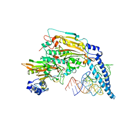
THE 2.9 ANGSTROMS CRYSTAL STRUCTURE OF T. THERMOPHILUS SERYL-TRNA SYNTHETASE COMPLEXED WITH TRNA SER
分子名称: PROTEIN (SERYL-TRNA SYNTHETASE (E.C.6.1.1.11)), TRNASER
著者Biou, S, Cusack, V, Yaremchuk, A, Tukalo, M.
登録日1994-02-21
公開日1994-04-30
最終更新日2024-02-14
実験手法X-RAY DIFFRACTION (2.9 Å)
主引用文献The 2.9 A crystal structure of T. thermophilus seryl-tRNA synthetase complexed with tRNA(Ser).
Science, 263, 1994
1H4A
DownloadVisualize
BU of 1h4a by Molmil
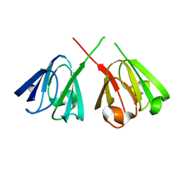
Human GammaD Crystallin R58H mutant structure AT 1.15 A resolution
分子名称: Gamma-crystallin D
著者Basak, A.K, Slingsby, C.
登録日2003-02-25
公開日2003-05-08
最終更新日2023-12-13
実験手法X-RAY DIFFRACTION (1.15 Å)
主引用文献High-Resolution X-Ray Crystal Structures of Human GammaD Crystallin (1.25A) and the R58H Mutant (1.15A) Associated with Aculeiform Cataract
J.Mol.Biol., 328, 2003
2BXJ
DownloadVisualize
BU of 2bxj by Molmil
Double Mutant of the Ribosomal Protein S6
分子名称: 30S RIBOSOMAL PROTEIN S6
著者Otzen, D.E.
登録日2005-07-26
公開日2005-10-26
最終更新日2023-12-13
実験手法X-RAY DIFFRACTION (2.4 Å)
主引用文献Antagonism, Non-Native Interactions and Non-Two-State Folding in S6 Revealed by Double-Mutant Cycle Analysis.
Protein Eng.Des.Sel., 18, 2005
<123


227111

件を2024-11-06に公開中

PDB statisticsPDBj update infoContact PDBjnumon