Loading
PDBj
メニューPDBj@FacebookPDBj@X(formerly Twitter)PDBj@BlueSkyPDBj@YouTubewwPDB FoundationwwPDB
RCSB PDBPDBeBMRBAdv. SearchSearch help
Search by PDB author
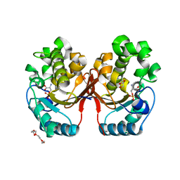
6A9C
DownloadVisualize
BU of 6a9c by Molmil
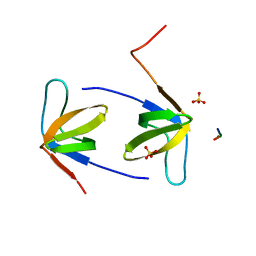
Crystal Structure c-terminal SH3 domain of Myosin IB from Entamoeba histolytica bound to EhFP10(GEF) peptide.
分子名称: Peptide from Rho guanine nucleotide exchange factor, SULFATE ION, Unconventional myosin IB
著者Gautam, G, Gourinath, S.
登録日2018-07-13
公開日2019-06-12
最終更新日2023-11-22
実験手法X-RAY DIFFRACTION (1.98 Å)
主引用文献EhFP10: A FYVE family GEF interacts with myosin IB to regulate cytoskeletal dynamics during endocytosis in Entamoeba histolytica.
Plos Pathog., 15, 2019
5ZR2
DownloadVisualize
BU of 5zr2 by Molmil
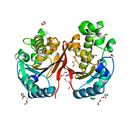
Crystal Structure of Phosphoserine Phosphatase Mutant (H9A) from Entamoeba histolytica in complex with Phosphoserine
分子名称: DI(HYDROXYETHYL)ETHER, PHOSPHOSERINE, Phosphoglycerate mutase family protein, ...
著者Kumari, P, Gourinath, S.
登録日2018-04-23
公開日2019-04-24
最終更新日2023-11-22
実験手法X-RAY DIFFRACTION (2.95 Å)
主引用文献Structural and functional characterisation of phosphoserine phosphatase, that plays critical role in the oxidative stress response in the parasite Entamoeba histolytica.
J.Struct.Biol., 206, 2019
5ZKK
DownloadVisualize
BU of 5zkk by Molmil
Crystal structure of Phosphoserine phosphatase from Entamoeba histolytica
分子名称: BETA-MERCAPTOETHANOL, DI(HYDROXYETHYL)ETHER, GLYCEROL, ...
著者Kumari, P, Gourinath, S.
登録日2018-03-24
公開日2019-04-03
最終更新日2023-11-22
実験手法X-RAY DIFFRACTION (2 Å)
主引用文献Structural and functional characterisation of phosphoserine phosphatase, that plays critical role in the oxidative stress response in the parasite Entamoeba histolytica.
J.Struct.Biol., 206, 2019
4LLB
DownloadVisualize
BU of 4llb by Molmil
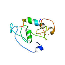
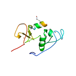
Crystal Structure of MOZ double PHD finger histone H3K14ac complex
分子名称: Histone H3.1, Histone acetyltransferase KAT6A, ZINC ION
著者Dreveny, I, Deeves, S.E, Yue, B, Heery, D.M.
登録日2013-07-09
公開日2013-10-16
最終更新日2024-10-30
実験手法X-RAY DIFFRACTION (2.5 Å)
主引用文献The double PHD finger domain of MOZ/MYST3 induces alpha-helical structure of the histone H3 tail to facilitate acetylation and methylation sampling and modification.
Nucleic Acids Res., 42, 2014
4LJN
DownloadVisualize
BU of 4ljn by Molmil
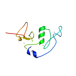
Crystal Structure of MOZ double PHD finger
分子名称: Histone acetyltransferase KAT6A, ZINC ION
著者Dreveny, I, Deeves, S.E, Yue, B, Heery, D.M.
登録日2013-07-05
公開日2013-10-16
最終更新日2023-09-20
実験手法X-RAY DIFFRACTION (3 Å)
主引用文献The double PHD finger domain of MOZ/MYST3 induces alpha-helical structure of the histone H3 tail to facilitate acetylation and methylation sampling and modification.
Nucleic Acids Res., 42, 2014
4LK9
DownloadVisualize
BU of 4lk9 by Molmil
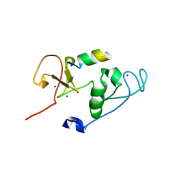
Crystal Structure of MOZ double PHD finger histone H3 tail complex
分子名称: Histone H3.1, Histone acetyltransferase KAT6A, ZINC ION
著者Dreveny, I, Deeves, S.E, Yue, B, Heery, D.M.
登録日2013-07-07
公開日2013-10-16
最終更新日2024-02-28
実験手法X-RAY DIFFRACTION (1.6 Å)
主引用文献The double PHD finger domain of MOZ/MYST3 induces alpha-helical structure of the histone H3 tail to facilitate acetylation and methylation sampling and modification.
Nucleic Acids Res., 42, 2014
4LKA
DownloadVisualize
BU of 4lka by Molmil
Crystal Structure of MOZ double PHD finger histone H3K9ac complex
分子名称: Histone H3.1, Histone acetyltransferase KAT6A, ZINC ION
著者Dreveny, I, Deeves, S.E, Yue, B, Heery, D.M.
登録日2013-07-07
公開日2013-10-16
最終更新日2023-12-06
実験手法X-RAY DIFFRACTION (1.61 Å)
主引用文献The double PHD finger domain of MOZ/MYST3 induces alpha-helical structure of the histone H3 tail to facilitate acetylation and methylation sampling and modification.
Nucleic Acids Res., 42, 2014
1YWL
DownloadVisualize
BU of 1ywl by Molmil
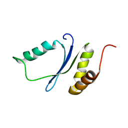
Solution NMR structure of the protein EF2693 from E. faecalis: Northeast Structural Genomics Consortium target EFR36
分子名称: Hypothetical UPF0213 protein EF2693
著者Northeast Structural Genomics Consortium (NESG)
登録日2005-02-18
公開日2005-03-29
最終更新日2024-05-22
実験手法SOLUTION NMR
主引用文献Solution NMR structure of the protein EF2693 from E. faecalis: Northeast Structural Genomics Consortium target EFR36
To be Published
2KOY
DownloadVisualize
BU of 2koy by Molmil
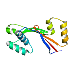
Structure of the E1064A mutant of the N-domain of Wilson Disease Associated Protein
分子名称: Copper-transporting ATPase 2
著者Dmitriev, O.Y.
登録日2009-10-03
公開日2010-12-15
最終更新日2024-05-22
実験手法SOLUTION NMR
主引用文献Difference in stability of the N-domain underlies distinct intracellular properties of the E1064A and H1069Q mutants of copper-transporting ATPase ATP7B.
J. Biol. Chem., 286, 2011
<123


227344

件を2024-11-13に公開中

PDB statisticsPDBj update infoContact PDBjnumon