6LV3
 
 | |
6LV7
 | |
6LUX
 | |
6LV1
 | |
6LVA
 | |
6LUW
 | |
6LV2
 | |
5Y2R
 | |
5Y2S
 | |
3TCX
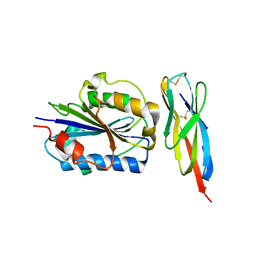 | | Structure of Engineered Single Domain ICAM-1 D1 with High-Affinity aL Integrin I Domain of Native C-Terminal Helix Conformation | | 分子名称: | Integrin alpha-L, Intercellular adhesion molecule 1, MAGNESIUM ION | | 著者 | Kang, S, Kim, C.U, Gu, X, Owens, R.M, van Rijn, S.J, Boonyaleepun, V, Mao, Y, Springer, T.A, Jin, M.M. | | 登録日 | 2011-08-09 | | 公開日 | 2011-08-31 | | 最終更新日 | 2014-11-12 | | 実験手法 | X-RAY DIFFRACTION (3.6 Å) | | 主引用文献 | Structure of Engineered Single Domain ICAM-1 D1 with High-Affinity L Integrin I Domain of Native C-Terminal Helix Conformation
To be Published
|
|
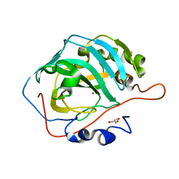
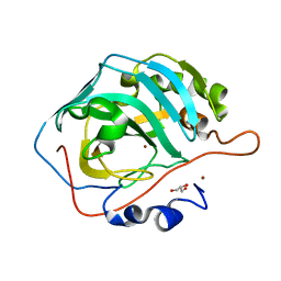
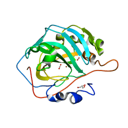
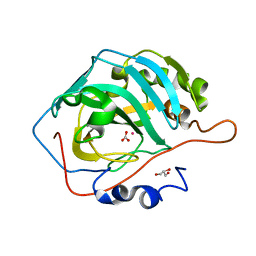
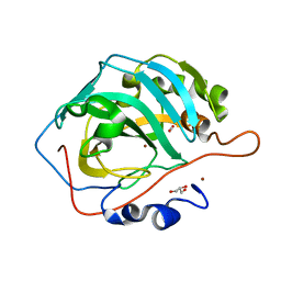
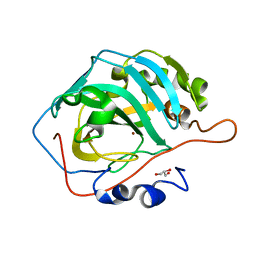
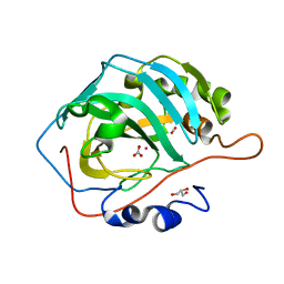
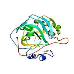
3U7C
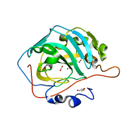
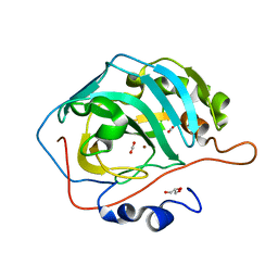 | | crystal structure of the V143I mutant of human carbonic anhydrase II | | 分子名称: | BICARBONATE ION, CARBON DIOXIDE, Carbonic anhydrase 2, ... | | 著者 | West, D.M, Kim, C.U, Robbins, A.H, Mckenna, R. | | 登録日 | 2011-10-13 | | 公開日 | 2013-02-13 | | 最終更新日 | 2023-09-13 | | 実験手法 | X-RAY DIFFRACTION (0.93 Å) | | 主引用文献 | crystal structure of the V143I mutant of human carbonic anhydrase II
To be Published
|
|
3DQ1
 | |
3DQA
 | |
3DQH
 | |
3DQ3
 | |
3DQD
 | |
3DQN
 | |
3DPX
 | |
3DQ5
 | |
3DQE
 | |
3DQJ
 | |
3DQI
 | |
3DQU
 | |
3DQ9
 | |
3DQM
 | |


