Loading
PDBj
メニューPDBj@FacebookPDBj@X(formerly Twitter)PDBj@BlueSkyPDBj@YouTubewwPDB FoundationwwPDBDonate
RCSB PDBPDBeBMRBAdv. SearchSearch help
Search by PDB author
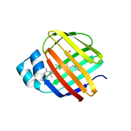
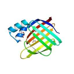
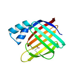
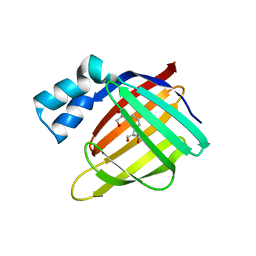
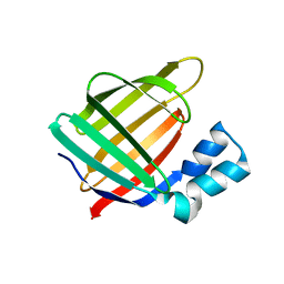
4EEJ
DownloadVisualize
BU of 4eej by Molmil
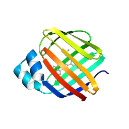
Crystal Structure of the Q108K:K40L:T51V:T53C:Y19W:R58W:T29L:Q4R Mutant of Cellular Retinol Binding Protein Type II in Complex with All-trans-Retinal at 1.5 Angstrom Resolution
分子名称: ACETATE ION, RETINAL, Retinol-binding protein 2
著者Nossoni, Z, Geiger, J.H.
登録日2012-03-28
公開日2012-12-26
最終更新日2024-10-30
実験手法X-RAY DIFFRACTION (1.501 Å)
主引用文献Tuning the electronic absorption of protein-embedded all-trans-retinal.
Science, 338, 2012
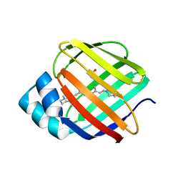
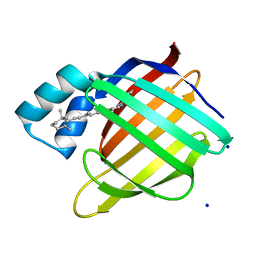
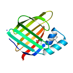
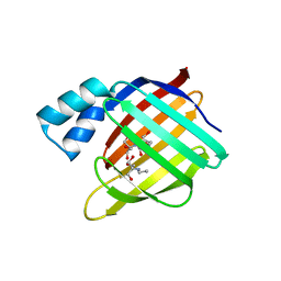
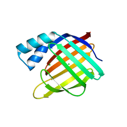
4GKC
DownloadVisualize
BU of 4gkc by Molmil
Crystal structure of Q108K:K40L:T51V:T53C:R58W:T29L:Y19W:Q4A mutant of cellular retinol binding protein II complex with all-trans-retinal at 1.33
分子名称: RETINAL, Retinol-binding protein 2
著者Nossoni, Z, Geiger, J.H.
登録日2012-08-10
公開日2012-12-26
最終更新日2024-11-20
実験手法X-RAY DIFFRACTION (1.3 Å)
主引用文献Tuning the electronic absorption of protein-embedded all-trans-retinal.
Science, 338, 2012
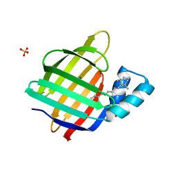
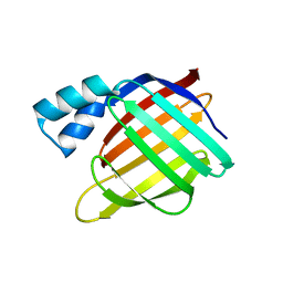
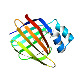
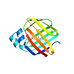
4EFG
DownloadVisualize
BU of 4efg by Molmil
Crystal Structure of the Q108K:K40L:T51V:T53C:Y19W:R58W:T29L Mutant of Cellular Retinol Binding Protein Type II in Complex with All-trans-Retinal at 1.58 Angstrom Resolution
分子名称: ACETATE ION, RETINAL, Retinol-binding protein 2
著者Nossoni, Z, Geiger, J.H.
登録日2012-03-29
公開日2012-12-26
最終更新日2024-10-16
実験手法X-RAY DIFFRACTION (1.58 Å)
主引用文献Tuning the electronic absorption of protein-embedded all-trans-retinal.
Science, 338, 2012
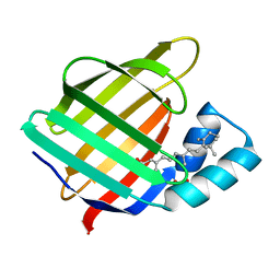
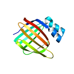
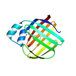
4EXZ
DownloadVisualize
BU of 4exz by Molmil
Crystal Structure of the Q108K:K40L Mutant of Cellular Retinol Binding Protein Type II in Complex with All-trans-Retinal at 1.7 Angstrom Resolution
分子名称: ACETATE ION, RETINAL, Retinol-binding protein 2
著者Nossoni, Z, Geiger, J.H.
登録日2012-05-01
公開日2012-12-26
最終更新日2024-11-06
実験手法X-RAY DIFFRACTION (1.608 Å)
主引用文献Tuning the electronic absorption of protein-embedded all-trans-retinal.
Science, 338, 2012
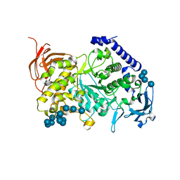
7ML5
DownloadVisualize
BU of 7ml5 by Molmil
Structure of the Starch Branching Enzyme I (BEI) complexed with maltododecaose from Oryza sativa L
分子名称: Isoform 2 of 1,4-alpha-glucan-branching enzyme, chloroplastic/amyloplastic, alpha-D-glucopyranose-(1-4)-alpha-D-glucopyranose-(1-4)-alpha-D-glucopyranose-(1-4)-alpha-D-glucopyranose, ...
著者Nayebi Gavgani, H, Fawaz, R, Geiger, J.H.
登録日2021-04-27
公開日2021-11-17
最終更新日2023-10-18
実験手法X-RAY DIFFRACTION (2.35 Å)
主引用文献A structural explanation for the mechanism and specificity of plant branching enzymes I and IIb.
J.Biol.Chem., 298, 2021
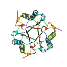
7MS9
DownloadVisualize
BU of 7ms9 by Molmil
Crystal structure of E114D mutant of Cg10062 with a covalent intermediate of the hydration of acetylenecarboxylic acid
分子名称: 3-HYDROXY-PROPANOIC ACID, 4-oxalocrotonate tautomerase, SULFATE ION
著者Nayebi, G.H, Geiger, J.H, Draths, K.
登録日2021-05-10
公開日2022-02-02
最終更新日2024-10-16
実験手法X-RAY DIFFRACTION (2.2 Å)
主引用文献Cg10062 Catalysis Forges a Link between Acetylenecarboxylic Acid and Bacterial Metabolism.
Biochemistry, 60, 2021
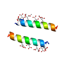
2DPQ
DownloadVisualize
BU of 2dpq by Molmil
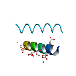
The crystal structures of the calcium-bound con-G and con-T(K7gamma) dimeric peptides demonstrate a novel metal-dependent helix-forming motif
分子名称: CALCIUM ION, CHLORIDE ION, Conantokin-G
著者Cnudde, S.E, Prorok, M, Dai, Q, Castellino, F.J, Geiger, J.H.
登録日2006-05-13
公開日2007-04-24
最終更新日2024-04-03
実験手法X-RAY DIFFRACTION (1.25 Å)
主引用文献The crystal structures of the calcium-bound con-G and con-T[K7gamma] dimeric peptides demonstrate a metal-dependent helix-forming motif
J.Am.Chem.Soc., 129, 2007
2DOI
DownloadVisualize
BU of 2doi by Molmil
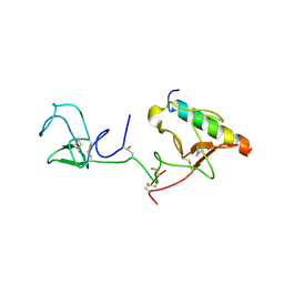
The X-ray crystallographic structure of the angiogenesis inhibitor, angiostatin, bound to a peptide from the group A streptococcus protein PAM
分子名称: Angiostatin, Plasminogen-binding group A streptococcal M-like protein PAM
著者Cnudde, S.E, Prorok, M, Castellino, F.J, Geiger, J.H.
登録日2006-04-29
公開日2006-12-05
最終更新日2024-10-16
実験手法X-RAY DIFFRACTION (3.1 Å)
主引用文献X-ray crystallographic structure of the angiogenesis inhibitor, angiostatin, bound to a peptide from the group A streptococcal surface protein PAM
Biochemistry, 45, 2006
2DOH
DownloadVisualize
BU of 2doh by Molmil
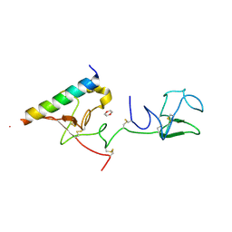
The X-ray crystallographic structure of the angiogenesis inhibitor, angiostatin, bound a to a peptide from the group A streptococcal surface protein PAM
分子名称: 1,4-DIETHYLENE DIOXIDE, Angiostatin, Plasminogen-binding group A streptococcal M-like protein PAM
著者Cnudde, S.E, Prorok, M, Castellino, F.J, Geiger, J.H.
登録日2006-04-29
公開日2006-12-05
最終更新日2024-10-09
実験手法X-RAY DIFFRACTION (2.3 Å)
主引用文献X-ray crystallographic structure of the angiogenesis inhibitor, angiostatin, bound to a peptide from the group A streptococcal surface protein PAM
Biochemistry, 45, 2006
2FRS
DownloadVisualize
BU of 2frs by Molmil
Crystal structure of the f15w mutant of apo-cellular retinoic acid binding protein type ii at 1.51 angstroms resolution
分子名称: Cellular retinoic acid-binding protein 2, SODIUM ION
著者Vaezeslami, S, Geiger, J.H.
登録日2006-01-20
公開日2006-09-19
最終更新日2024-02-14
実験手法X-RAY DIFFRACTION (1.51 Å)
主引用文献The structure of Apo-wild-type cellular retinoic acid binding protein II at 1.4 A and its relationship to ligand binding and nuclear translocation.
J.Mol.Biol., 363, 2006
6C7Z
DownloadVisualize
BU of 6c7z by Molmil
Crystal structure of the Q108K:K40L:T51V:R58F mutant of human Cellular Retinol Binding Protein II in complex with synthetic Ligand Julolidine
分子名称: (2E,4E)-3-methyl-5-(2,3,6,7-tetrahydro-1H,5H-pyrido[3,2,1-ij]quinolin-9-yl)penta-2,4-dienal, ACETATE ION, Retinol-binding protein 2
著者Nosrati, M, Geiger, J.H.
登録日2018-01-23
公開日2018-04-25
最終更新日2024-11-13
実験手法X-RAY DIFFRACTION (1.42 Å)
主引用文献A Genetically Encoded Ratiometric pH Probe: Wavelength Regulation-Inspired Design of pH Indicators.
Chembiochem, 19, 2018
2FS6
DownloadVisualize
BU of 2fs6 by Molmil
Crystal Structure of Apo-Cellular Retinoic Acid Binding Protein Type II At 1.35 Angstroms Resolution
分子名称: ACETATE ION, CHLORIDE ION, Cellular retinoic acid-binding protein 2, ...
著者Vaezeslami, S, Geiger, J.H.
登録日2006-01-21
公開日2006-09-19
最終更新日2024-02-14
実験手法X-RAY DIFFRACTION (1.35 Å)
主引用文献The structure of Apo-wild-type cellular retinoic acid binding protein II at 1.4 A and its relationship to ligand binding and nuclear translocation.
J.Mol.Biol., 363, 2006
2G7B
DownloadVisualize
BU of 2g7b by Molmil
Crystal Structure of the R132K:R111L:L121E mutant of Cellular Retinoic Acid Binding Protein Type II In Complex With All-Trans-Retinal At 1.18 Angstroms Resolution
分子名称: Cellular retinoic acid-binding protein 2, RETINAL, SODIUM ION
著者Vaezeslami, S, Geiger, J.H.
登録日2006-02-27
公開日2007-04-17
最終更新日2025-01-29
実験手法X-RAY DIFFRACTION (1.18 Å)
主引用文献Protein design: reengineering cellular retinoic acid binding protein II into a rhodopsin protein mimic.
J.Am.Chem.Soc., 129, 2007
2G79
DownloadVisualize
BU of 2g79 by Molmil
Crystal Structure of the R132K:Y134F Mutant of Cellular Retinoic Acid Binding Protein Type II in Complex with All-Trans-Retinal at 1.69 Angstroms Resolution
分子名称: Cellular retinoic acid-binding protein 2, RETINAL, SODIUM ION, ...
著者Vaezeslami, S, Geiger, J.H.
登録日2006-02-27
公開日2007-04-17
最終更新日2024-02-14
実験手法X-RAY DIFFRACTION (1.69 Å)
主引用文献Protein design: reengineering cellular retinoic acid binding protein II into a rhodopsin protein mimic.
J.Am.Chem.Soc., 129, 2007
2FR3
DownloadVisualize
BU of 2fr3 by Molmil
Crystal Structure of Cellular Retinoic Acid Binding Protein Type II in Complex with All-Trans-Retinoic Acid at 1.48 Angstroms Resolution
分子名称: ACETATE ION, Cellular retinoic acid binding protein 2, RETINOIC ACID
著者Vaezeslami, S, Geiger, J.H.
登録日2006-01-18
公開日2006-09-19
最終更新日2024-02-14
実験手法X-RAY DIFFRACTION (1.48 Å)
主引用文献The structure of Apo-wild-type cellular retinoic acid binding protein II at 1.4 A and its relationship to ligand binding and nuclear translocation.
J.Mol.Biol., 363, 2006
2FS7
DownloadVisualize
BU of 2fs7 by Molmil
Crystal Structure of Apo-Cellular Retinoic Acid Binding Protein Type II At 1.55 Angstroms Resolution
分子名称: ACETATE ION, CHLORIDE ION, Cellular retinoic acid-binding protein 2
著者Vaezeslami, S, Geiger, J.H.
登録日2006-01-21
公開日2006-09-19
最終更新日2024-02-14
実験手法X-RAY DIFFRACTION (1.55 Å)
主引用文献The structure of Apo-wild-type cellular retinoic acid binding protein II at 1.4 A and its relationship to ligand binding and nuclear translocation.
J.Mol.Biol., 363, 2006
2G78
DownloadVisualize
BU of 2g78 by Molmil
Crystal Structure of the R132K:Y134F Mutant of Cellular Retinoic Acid Binding Protein Type II in Complex with All-Trans-Retinoic Acid at 1.70 Angstroms Resolution
分子名称: Cellular retinoic acid-binding protein 2, RETINOIC ACID, SODIUM ION
著者Vaezeslami, S, Geiger, J.H.
登録日2006-02-27
公開日2007-04-17
最終更新日2024-02-14
実験手法X-RAY DIFFRACTION (1.7 Å)
主引用文献Protein design: reengineering cellular retinoic acid binding protein II into a rhodopsin protein mimic.
J.Am.Chem.Soc., 129, 2007
2DPR
DownloadVisualize
BU of 2dpr by Molmil
The crystal structures of the calcium-bound con-G and con-T(K7Glu) dimeric peptides demonstrate a novel metal-dependent helix-forming motif
分子名称: CALCIUM ION, Conantokin-T
著者Cnudde, S.E, Prorok, M, Dai, Q, Castellino, F.J, Geiger, J.H.
登録日2006-05-13
公開日2007-04-24
最終更新日2024-04-03
実験手法X-RAY DIFFRACTION (1.7 Å)
主引用文献The crystal structures of the calcium-bound con-G and con-T[K7gamma] dimeric peptides demonstrate a metal-dependent helix-forming motif
J.Am.Chem.Soc., 129, 2007
3D95
DownloadVisualize
BU of 3d95 by Molmil
Crystal Structure of the R132K:Y134F:R111L:L121E:T54V Mutant of Apo-Cellular Retinoic Acid Binding Protein Type II at 1.20 Angstroms Resolution
分子名称: Cellular retinoic acid-binding protein 2
著者Vaezeslami, S, Geiger, J.H.
登録日2008-05-26
公開日2008-07-22
最終更新日2024-02-21
実験手法X-RAY DIFFRACTION (1.2 Å)
主引用文献Structural analysis of site-directed mutants of cellular retinoic acid-binding protein II addresses the relationship between structural integrity and ligand binding.
Acta Crystallogr.,Sect.D, 64, 2008
3D97
DownloadVisualize
BU of 3d97 by Molmil
Crystal Structure of the R132K:R111L:L121E Mutant of Apo-Cellular Retinoic Acid Binding Protein Type II At 1.50 Angstroms Resolution
分子名称: 2-[3-(2-HYDROXY-1,1-DIHYDROXYMETHYL-ETHYLAMINO)-PROPYLAMINO]-2-HYDROXYMETHYL-PROPANE-1,3-DIOL, Cellular retinoic acid-binding protein 2, SODIUM ION
著者Vaezeslami, S, Geiger, J.H.
登録日2008-05-26
公開日2008-07-22
最終更新日2023-08-30
実験手法X-RAY DIFFRACTION (1.5 Å)
主引用文献Determining Crystal Structures of Proteins and Protein Complexes by X-Ray Crystallography: X-Ray Crystallographic Studies of the Mutants of Cellular Retinoic Acid Binding Protein Type II Toward Designing a Mimic of Rhodopsin.
Thesis
3FEL
DownloadVisualize
BU of 3fel by Molmil
Crystal structure of the R132K:R111L:T54E mutant of cellular retinoic acid-binding protein II at 1.85 angstrom resolution
分子名称: 2-[3-(2-HYDROXY-1,1-DIHYDROXYMETHYL-ETHYLAMINO)-PROPYLAMINO]-2-HYDROXYMETHYL-PROPANE-1,3-DIOL, Cellular retinoic acid-binding protein 2
著者Jia, X, Geiger, J.H.
登録日2008-11-30
公開日2009-11-10
最終更新日2023-09-06
実験手法X-RAY DIFFRACTION (1.85 Å)
主引用文献Crystal structures of apo cellular retinoic acid-binding protein II mutants: Structural integrity investigated through multiple site mutations
To be Published
3FEP
DownloadVisualize
BU of 3fep by Molmil
Crystal structure of the R132K:R111L:L121E:R59W-CRABPII mutant complexed with a synthetic ligand (merocyanin) at 2.60 angstrom resolution.
分子名称: (2E,4E,6E)-3-methyl-6-(1,3,3-trimethyl-1,3-dihydro-2H-indol-2-ylidene)hexa-2,4-dienal, 2-(N-MORPHOLINO)-ETHANESULFONIC ACID, Cellular retinoic acid-binding protein 2
著者Jia, X, Geiger, J.H.
登録日2008-11-30
公開日2009-11-10
最終更新日2024-10-16
実験手法X-RAY DIFFRACTION (2.6 Å)
主引用文献"Turn-on" protein fluorescence: in situ formation of cyanine dyes.
J.Am.Chem.Soc., 137, 2015
3FA9
DownloadVisualize
BU of 3fa9 by Molmil
Crystal structure of the apo R132K:Y134F:R111L:L121D mutant of cellular retinoic acid-binding protein II at 1.94 angstrom resolution
分子名称: ACETATE ION, Cellular retinoic acid-binding protein 2
著者Jia, X, Geiger, J.H.
登録日2008-11-16
公開日2009-10-27
最終更新日2023-12-27
実験手法X-RAY DIFFRACTION (1.94 Å)
主引用文献Crystal structures of apo cellular retinoic acid-binding protein II mutants: Structural integrity investigated through multiple site mutations
To be Published
3FA8
DownloadVisualize
BU of 3fa8 by Molmil
Crystal structure of the apo R132K:Y134F:R111L:L121E mutant of cellular retinoic acid-binding protein II at 1.78 angstrom resolution
分子名称: Cellular retinoic acid-binding protein 2
著者Jia, X, Geiger, J.H.
登録日2008-11-16
公開日2009-10-27
最終更新日2023-09-06
実験手法X-RAY DIFFRACTION (1.78 Å)
主引用文献Crystal structures of apo cellular retinoic acid-binding protein II mutants: Structural integrity investigated through multiple site mutations
To be Published
3FEN
DownloadVisualize
BU of 3fen by Molmil
Crystal structure of the R132K:R111L:A32E mutant of cellular retinoic acid-binding protein II at 1.56 angstrom resolution
分子名称: CHLORIDE ION, Cellular retinoic acid-binding protein 2
著者Jia, X, Geiger, J.H.
登録日2008-11-30
公開日2009-11-10
最終更新日2023-09-06
実験手法X-RAY DIFFRACTION (1.56 Å)
主引用文献Crystal structures of apo cellular retinoic acid-binding protein II mutants: Structural integrity investigated through multiple site mutations
To be Published

238582

件を2025-07-09に公開中

PDB statisticsPDBj update infoContact PDBjnumon