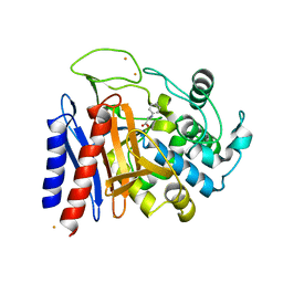
2CU1
 
 | |
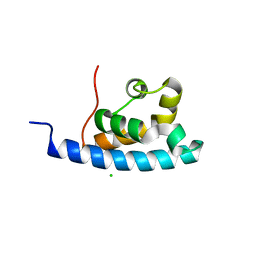
2ENQ
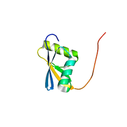
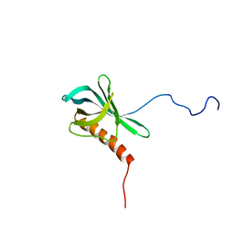
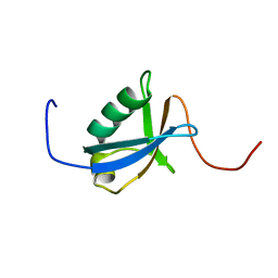
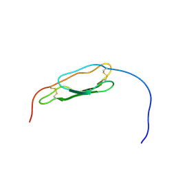
 | | Solution structure of the C2 domain from human PI3-kinase p110 subunit alpha | | 分子名称: | Phosphatidylinositol-4,5-bisphosphate 3-kinase catalytic subunit alpha isoform | | 著者 | Endo, H, Nagashima, T, Yoshida, M, Hayashi, F, Yokoyama, S, RIKEN Structural Genomics/Proteomics Initiative (RSGI) | | 登録日 | 2007-03-28 | | 公開日 | 2008-04-01 | | 最終更新日 | 2024-05-29 | | 実験手法 | SOLUTION NMR | | 主引用文献 | Solution structure of the C2 domain from human PI3-kinase p110 subunit alpha
To be Published
|
|
2EOB
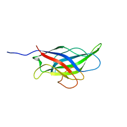
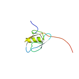
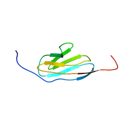
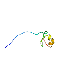
 | | Solution structure of the second SH2 domain from rat PLC gamma-2 | | 分子名称: | 1-phosphatidylinositol-4,5-bisphosphate phosphodiesterase gamma 2 | | 著者 | Sano, R, Hayashi, F, Nagashima, T, Yoshida, M, Yokoyama, S, RIKEN Structural Genomics/Proteomics Initiative (RSGI) | | 登録日 | 2007-03-29 | | 公開日 | 2008-04-01 | | 最終更新日 | 2024-05-29 | | 実験手法 | SOLUTION NMR | | 主引用文献 | Solution structure of the second SH2 domain from rat PLC gamma-2
To be Published
|
|
2EQI
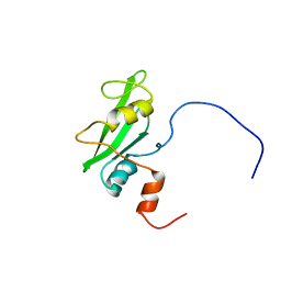
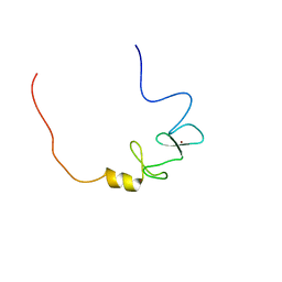
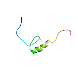
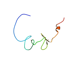
 | | Solution structure of the SH3 domain from Phospholipase C, gamma 2 | | 分子名称: | Phospholipase C, gamma 2 | | 著者 | Qin, X.R, Nagashima, T, Hayahsi, F, Yokoyama, S, RIKEN Structural Genomics/Proteomics Initiative (RSGI) | | 登録日 | 2007-03-30 | | 公開日 | 2008-04-08 | | 最終更新日 | 2024-05-29 | | 実験手法 | SOLUTION NMR | | 主引用文献 | Solution structure of the SH3 domain from Phospholipase C, gamma 2
To be Published
|
|
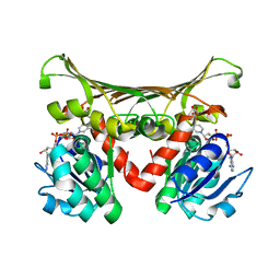
2CRF
 | |
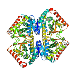
2D90
 | |
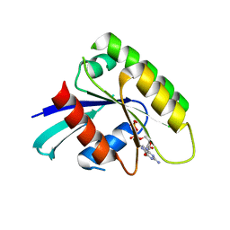
2D8Y
 | |
2DNF
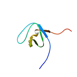
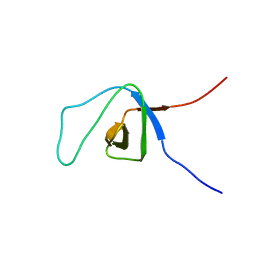
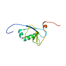 | | Solution structure of RSGI RUH-062, a DCX domain from human | | 分子名称: | Doublecortin domain-containing protein 2 | | 著者 | Ohashi, W, Hirota, H, Nagashima, T, Hayashi, F, Yokoyama, S, RIKEN Structural Genomics/Proteomics Initiative (RSGI) | | 登録日 | 2006-04-26 | | 公開日 | 2006-10-26 | | 最終更新日 | 2024-05-29 | | 実験手法 | SOLUTION NMR | | 主引用文献 | Solution structure of RSGI RUH-062, a DCX domain from human
TO BE PUBLISHED
|
|
2EEL
 | |
2EL4
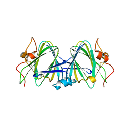
 | | Solution structure of the 15th zf-C2H2 domain from human Zinc finger protein 268 | | 分子名称: | ZINC ION, Zinc finger protein 268 | | 著者 | Kurosaki, C, Nagashima, T, Yoshida, M, Hayashi, F, Yokoyama, S, RIKEN Structural Genomics/Proteomics Initiative (RSGI) | | 登録日 | 2007-03-26 | | 公開日 | 2007-10-02 | | 最終更新日 | 2024-05-01 | | 実験手法 | SOLUTION NMR | | 主引用文献 | Solution structure of the 15th zf-C2H2 domain from human Zinc finger protein 268
To be Published
|
|
2E7B
 | |
2EGE
 | |
2EHF
 | |
2EGM
 | |
2EHE
 | |
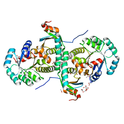
2EMS
 | | Crystal Structure Analysis of the radixin FERM domain complexed with adhesion molecule CD43 | | 分子名称: | Leukosialin, Radixin | | 著者 | Takai, Y, Kitano, K, Terawaki, S, Maesaki, R, Hakoshima, T. | | 登録日 | 2007-03-28 | | 公開日 | 2008-04-01 | | 最終更新日 | 2023-10-25 | | 実験手法 | X-RAY DIFFRACTION (2.9 Å) | | 主引用文献 | Structural basis of the cytoplasmic tail of adhesion molecule CD43 and its binding to ERM proteins
J.Mol.Biol., 381, 2008
|
|
2EMT
 | | Crystal Structure Analysis of the radixin FERM domain complexed with adhesion molecule PSGL-1 | | 分子名称: | P-selectin glycoprotein ligand 1, Radixin | | 著者 | Takai, Y, Kitano, K, Terawaki, S, Maesaki, R, Hakoshima, T. | | 登録日 | 2007-03-28 | | 公開日 | 2008-03-18 | | 最終更新日 | 2023-10-25 | | 実験手法 | X-RAY DIFFRACTION (2.8 Å) | | 主引用文献 | Structural basis of PSGL-1 binding to ERM proteins
Genes Cells, 12, 2007
|
|
2EFX
 | | The crystal structure of D-amino acid amidase from Ochrobactrum anthropi SV3 complexed with L-phenylalanine amide | | 分子名称: | BARIUM ION, D-amino acid amidase, PHENYLALANINE AMIDE | | 著者 | Okazaki, S, Suzuki, A, Mizushima, T, Komeda, H, Asano, Y, Yamane, T. | | 登録日 | 2007-02-26 | | 公開日 | 2007-03-06 | | 最終更新日 | 2023-10-25 | | 実験手法 | X-RAY DIFFRACTION (2.2 Å) | | 主引用文献 | Structures of D-amino-acid amidase complexed with L-phenylalanine and with L-phenylalanine amide: insight into the D-stereospecificity of D-amino-acid amidase from Ochrobactrum anthropi SV3.
Acta Crystallogr.,Sect.D, 64, 2008
|
|
2B9U
 | | Crystal structure of dTDP-4-dehydrorhamnose 3,5-epimerase from sulfolobus tokodaii | | 分子名称: | hypothetical dTDP-4-dehydrorhamnose 3,5-epimerase | | 著者 | Rajakannan, V, Kondo, K, Mizushima, T, Suzuki, A, Yamane, T. | | 登録日 | 2005-10-13 | | 公開日 | 2006-10-13 | | 最終更新日 | 2023-10-25 | | 実験手法 | X-RAY DIFFRACTION (2.07 Å) | | 主引用文献 | Crystal structure of dTDP-4-dehydrorhamnose 3,5-epimerase from sulfolobus tokodaii
TO BE PUBLISHED
|
|
2EFU
 | | The crystal structure of D-amino acid amidase from Ochrobactrum anthropi SV3 complexed with L-phenylalanine | | 分子名称: | BARIUM ION, D-Amino acid amidase, PHENYLALANINE | | 著者 | Okazaki, S, Suzuki, A, Mizushima, T, Komeda, H, Asano, Y, Yamane, T. | | 登録日 | 2007-02-26 | | 公開日 | 2007-03-06 | | 最終更新日 | 2023-10-25 | | 実験手法 | X-RAY DIFFRACTION (2.3 Å) | | 主引用文献 | Structures of D-amino-acid amidase complexed with L-phenylalanine and with L-phenylalanine amide: insight into the D-stereospecificity of D-amino-acid amidase from Ochrobactrum anthropi SV3.
Acta Crystallogr.,Sect.D, 64, 2008
|
|
2DC1
 | | Crystal Structure Of L-Aspartate Dehydrogenase From Hyperthermophilic Archaeon Archaeoglobus fulgidus | | 分子名称: | CITRIC ACID, L-aspartate dehydrogenase, NICOTINAMIDE-ADENINE-DINUCLEOTIDE | | 著者 | Yoneda, K, Sakuraba, H, Tsuge, H, Ohshima, T. | | 登録日 | 2005-12-19 | | 公開日 | 2006-12-26 | | 最終更新日 | 2024-03-13 | | 実験手法 | X-RAY DIFFRACTION (1.9 Å) | | 主引用文献 | Crystal structure of archaeal highly thermostable L-aspartate dehydrogenase/NAD/citrate ternary complex.
Febs J., 274, 2007
|
|
2D4A
 | | Structure of the malate dehydrogenase from Aeropyrum pernix | | 分子名称: | Malate dehydrogenase | | 著者 | Kawakami, R, Sakuraba, H, Tsuge, H, Ohshima, T. | | 登録日 | 2005-10-12 | | 公開日 | 2006-11-14 | | 最終更新日 | 2024-03-13 | | 実験手法 | X-RAY DIFFRACTION (2.87 Å) | | 主引用文献 | Refolding, characterization and crystal structure of (S)-malate dehydrogenase from the hyperthermophilic archaeon Aeropyrum pernix.
Biochim.Biophys.Acta, 1794, 2009
|
|
2DPX
 | | Crystal Structure of human Rad GTPase | | 分子名称: | GTP-binding protein RAD, GUANOSINE-5'-DIPHOSPHATE, MAGNESIUM ION | | 著者 | Yanuar, A, Sakurai, S, Kitano, K, Hakoshima, T. | | 登録日 | 2006-05-18 | | 公開日 | 2006-08-08 | | 最終更新日 | 2023-10-25 | | 実験手法 | X-RAY DIFFRACTION (1.8 Å) | | 主引用文献 | Crystal structure of human Rad GTPase of the RGK-family
Genes Cells, 11, 2006
|
|
2E1E
 | | Crystal structure of the HRDC Domain of Human Werner Syndrome Protein, WRN | | 分子名称: | CHLORIDE ION, Werner syndrome ATP-dependent helicase | | 著者 | Kitano, K, Yoshihara, N, Hakoshima, T. | | 登録日 | 2006-10-25 | | 公開日 | 2006-12-12 | | 最終更新日 | 2023-10-25 | | 実験手法 | X-RAY DIFFRACTION (2.3 Å) | | 主引用文献 | Crystal structure of the HRDC domain of human Werner syndrome protein, WRN
J.Biol.Chem., 282, 2007
|
|
2CZV
 | | Crystal structure of archeal RNase P protein ph1481p in complex with ph1877p | | 分子名称: | ACETIC ACID, Ribonuclease P protein component 2, Ribonuclease P protein component 3, ... | | 著者 | Kawano, S, Kakuta, Y, Nakashima, T, Tanaka, I, Kimura, M. | | 登録日 | 2005-07-19 | | 公開日 | 2006-06-27 | | 最終更新日 | 2024-05-29 | | 実験手法 | X-RAY DIFFRACTION (2 Å) | | 主引用文献 | Crystal structure of protein Ph1481p in complex with protein Ph1877p of archaeal RNase P from Pyrococcus horikoshii OT3: implication of dimer formation of the holoenzyme
J.Mol.Biol., 357, 2006
|
|


