1VGW
 
 | |
1VHV
 | |
1VIC
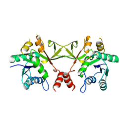
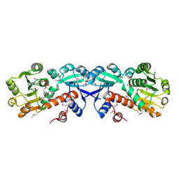
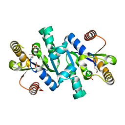
 | | Crystal structure of CMP-KDO synthetase | | 分子名称: | 3-deoxy-manno-octulosonate cytidylyltransferase | | 著者 | Structural GenomiX | | 登録日 | 2003-12-01 | | 公開日 | 2003-12-30 | | 最終更新日 | 2023-12-27 | | 実験手法 | X-RAY DIFFRACTION (1.8 Å) | | 主引用文献 | Structural analysis of a set of proteins resulting from a bacterial genomics project
Proteins, 60, 2005
|
|
1VIV
 | |
1VI2
 | |
1VIM
 | |
1VGX
 | |
1VH8
 | |
1VI3
 | |
1VIA
 | |
1VIQ
 | |
1VH4
 | |
1VHO
 | |
1VHY
 | |
1VIY
 | |
1VH7
 | |
1VHF
 | |
1VHZ
 | |
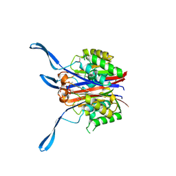
1VIS
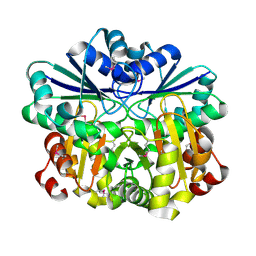
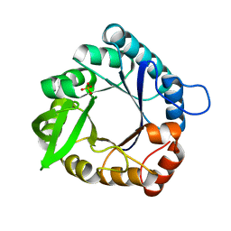
 | | Crystal structure of mevalonate kinase | | 分子名称: | 1,4-DIETHYLENE DIOXIDE, Mevalonate kinase | | 著者 | Structural GenomiX | | 登録日 | 2003-12-01 | | 公開日 | 2003-12-30 | | 最終更新日 | 2023-12-27 | | 実験手法 | X-RAY DIFFRACTION (2.69 Å) | | 主引用文献 | Structural analysis of a set of proteins resulting from a bacterial genomics project
Proteins, 60, 2005
|
|
1VIZ
 | |
1VHA
 | |
1VHU
 | |
1VI5
 | |
1VHG
 | |
1VHT
 | |


