Loading
PDBj
メニューPDBj@FacebookPDBj@TwitterPDBj@YouTubewwPDB FoundationwwPDB
RCSB PDBPDBeBMRBAdv. SearchSearch help
Search by PDB author
8HVX
DownloadVisualize
BU of 8hvx by Molmil
Crystal structure of SARS-Cov-2 main protease Y54C mutant in complex with PF07304814
分子名称: 3C-like proteinase nsp5, [(3~{S})-3-[[(2~{S})-2-[(4-methoxy-1~{H}-indol-2-yl)carbonylamino]-4-methyl-pentanoyl]amino]-2-oxidanylidene-4-[(3~{R})-2-oxidanylidene-3,4-dihydropyrrol-3-yl]butyl] dihydrogen phosphate
著者Zhou, X.L, Zhang, J, Li, J.
登録日2022-12-28
公開日2024-01-17
最終更新日2024-02-07
実験手法X-RAY DIFFRACTION (1.75 Å)
主引用文献Crystal structures of main protease (M pro ) mutants of SARS-CoV-2 variants bound to PF-07304814.
Mol Biomed, 4, 2023
8HVY
DownloadVisualize
BU of 8hvy by Molmil
Crystal structure of SARS-Cov-2 main protease K90R mutant in complex with PF07304814
分子名称: 3C-like proteinase nsp5, [(3~{S})-3-[[(2~{S})-2-[(4-methoxy-1~{H}-indol-2-yl)carbonylamino]-4-methyl-pentanoyl]amino]-2-oxidanylidene-4-[(3~{R})-2-oxidanylidene-3,4-dihydropyrrol-3-yl]butyl] dihydrogen phosphate
著者Wang, J, Zhang, J, Li, J.
登録日2022-12-28
公開日2024-01-17
最終更新日2024-02-07
実験手法X-RAY DIFFRACTION (1.97 Å)
主引用文献Crystal structures of main protease (M pro ) mutants of SARS-CoV-2 variants bound to PF-07304814.
Mol Biomed, 4, 2023
8HVZ
DownloadVisualize
BU of 8hvz by Molmil
Crystal structure of SARS-Cov-2 main protease V186F mutant in complex with PF07304814
分子名称: 3C-like proteinase nsp5, [(3~{S})-3-[[(2~{S})-2-[(4-methoxy-1~{H}-indol-2-yl)carbonylamino]-4-methyl-pentanoyl]amino]-2-oxidanylidene-4-[(3~{R})-2-oxidanylidene-3,4-dihydropyrrol-3-yl]butyl] dihydrogen phosphate
著者Zou, X.F, Zhang, J, Li, J.
登録日2022-12-28
公開日2024-01-17
最終更新日2024-02-07
実験手法X-RAY DIFFRACTION (1.7 Å)
主引用文献Crystal structures of main protease (M pro ) mutants of SARS-CoV-2 variants bound to PF-07304814.
Mol Biomed, 4, 2023
8HVW
DownloadVisualize
BU of 8hvw by Molmil
Crystal structure of SARS-Cov-2 main protease M49I mutant in complex with PF07304814
分子名称: 3C-like proteinase nsp5, [(3~{S})-3-[[(2~{S})-2-[(4-methoxy-1~{H}-indol-2-yl)carbonylamino]-4-methyl-pentanoyl]amino]-2-oxidanylidene-4-[(3~{R})-2-oxidanylidene-3,4-dihydropyrrol-3-yl]butyl] dihydrogen phosphate
著者Zou, X.F, Zhang, J, Li, J.
登録日2022-12-28
公開日2024-01-17
最終更新日2024-02-07
実験手法X-RAY DIFFRACTION (2.05 Å)
主引用文献Crystal structures of main protease (M pro ) mutants of SARS-CoV-2 variants bound to PF-07304814.
Mol Biomed, 4, 2023
8IG4
DownloadVisualize
BU of 8ig4 by Molmil
Crystal structure of SARS-Cov-2 main protease in complex with GC376
分子名称: Non-structural protein 11, N~2~-[(benzyloxy)carbonyl]-N-{(2S)-1-hydroxy-3-[(3S)-2-oxopyrrolidin-3-yl]propan-2-yl}-L-leucinamide
著者Zhou, X.L, Zhang, J, Li, J.
登録日2023-02-20
公開日2024-03-06
最終更新日2024-05-08
実験手法X-RAY DIFFRACTION (1.8 Å)
主引用文献Structural Basis for Coronaviral Main Proteases Inhibition by the 3CLpro Inhibitor GC376.
J.Mol.Biol., 436, 2024
8IG5
DownloadVisualize
BU of 8ig5 by Molmil
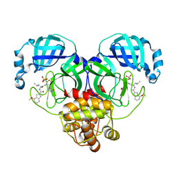
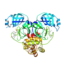
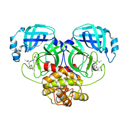
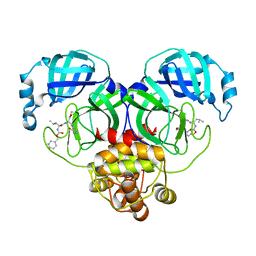
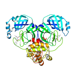
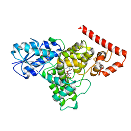
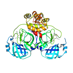
Crystal structure of SARS main protease in complex with GC376
分子名称: 3C-like proteinase nsp5, N~2~-[(benzyloxy)carbonyl]-N-{(2S)-1-hydroxy-3-[(3S)-2-oxopyrrolidin-3-yl]propan-2-yl}-L-leucinamide
著者Lin, C, Zhang, J, Li, J.
登録日2023-02-20
公開日2024-03-06
最終更新日2024-05-08
実験手法X-RAY DIFFRACTION (2.17 Å)
主引用文献Structural Basis for Coronaviral Main Proteases Inhibition by the 3CLpro Inhibitor GC376.
J.Mol.Biol., 436, 2024
8IG6
DownloadVisualize
BU of 8ig6 by Molmil
Crystal structure of MERS main protease in complex with GC376
分子名称: N~2~-[(benzyloxy)carbonyl]-N-{(2S)-1-hydroxy-3-[(3S)-2-oxopyrrolidin-3-yl]propan-2-yl}-L-leucinamide, ORF1a
著者Lin, C, Zhang, J, Li, J.
登録日2023-02-20
公開日2024-03-06
最終更新日2024-05-08
実験手法X-RAY DIFFRACTION (2.07 Å)
主引用文献Structural Basis for Coronaviral Main Proteases Inhibition by the 3CLpro Inhibitor GC376.
J.Mol.Biol., 436, 2024
8IG9
DownloadVisualize
BU of 8ig9 by Molmil
Crystal structure of SARS-Cov-2 main protease M49I mutant in complex with GC376
分子名称: 3C-like proteinase nsp5, N~2~-[(benzyloxy)carbonyl]-N-{(2S)-1-hydroxy-3-[(3S)-2-oxopyrrolidin-3-yl]propan-2-yl}-L-leucinamide
著者Zou, X.F, Zhang, J, Li, J.
登録日2023-02-20
公開日2024-03-06
最終更新日2024-05-08
実験手法X-RAY DIFFRACTION (1.68 Å)
主引用文献Structural Basis for Coronaviral Main Proteases Inhibition by the 3CLpro Inhibitor GC376.
J.Mol.Biol., 436, 2024
8IGA
DownloadVisualize
BU of 8iga by Molmil
Crystal structure of SARS-Cov-2 main protease K90R mutant in complex with GC376
分子名称: 3C-like proteinase nsp5, N~2~-[(benzyloxy)carbonyl]-N-{(2S)-1-hydroxy-3-[(3S)-2-oxopyrrolidin-3-yl]propan-2-yl}-L-leucinamide
著者Wang, J, Zhang, J, Li, J.
登録日2023-02-20
公開日2024-03-06
最終更新日2024-05-08
実験手法X-RAY DIFFRACTION (1.63 Å)
主引用文献Structural Basis for Coronaviral Main Proteases Inhibition by the 3CLpro Inhibitor GC376.
J.Mol.Biol., 436, 2024
8IGB
DownloadVisualize
BU of 8igb by Molmil
Crystal structure of SARS-Cov-2 main protease P132H mutant in complex with GC376
分子名称: 3C-like proteinase nsp5, N~2~-[(benzyloxy)carbonyl]-N-{(2S)-1-hydroxy-3-[(3S)-2-oxopyrrolidin-3-yl]propan-2-yl}-L-leucinamide
著者Li, W.W, Zhang, J, Li, J.
登録日2023-02-20
公開日2024-03-06
最終更新日2024-05-08
実験手法X-RAY DIFFRACTION (1.72 Å)
主引用文献Structural Basis for Coronaviral Main Proteases Inhibition by the 3CLpro Inhibitor GC376.
J.Mol.Biol., 436, 2024
5LFA
DownloadVisualize
BU of 5lfa by Molmil
Crystal structure of iron-sulfur cluster containing bacterial (6-4) photolyase PhrB - Y424F mutant with impaired DNA repair activity
分子名称: (6-4) photolyase, 1-deoxy-1-(6,7-dimethyl-2,4-dioxo-3,4-dihydropteridin-8(2H)-yl)-D-ribitol, FLAVIN-ADENINE DINUCLEOTIDE, ...
著者Kwiatkowski, D, Zhang, F, Krauss, N, Lamparter, T, Scheerer, P.
登録日2016-06-30
公開日2017-01-11
最終更新日2024-01-10
実験手法X-RAY DIFFRACTION (2.5 Å)
主引用文献Crystal Structures of Bacterial (6-4) Photolyase Mutants with Impaired DNA Repair Activity.
Photochem. Photobiol., 93, 2017
6CF8
DownloadVisualize
BU of 6cf8 by Molmil
Crystal structure of Cj0843 lytic transglycosylase of Campylobacter jejuni at 1.87A resolution
分子名称: Lytic transglycosylase, SULFATE ION
著者van den Akker, F, Kumar, V, Vijayaraghavan, J.
登録日2018-02-13
公開日2018-05-30
実験手法X-RAY DIFFRACTION (1.87 Å)
主引用文献Structural studies and molecular dynamics simulations suggest a processive mechanism of exolytic lytic transglycosylase from Campylobacter jejuni.
PLoS ONE, 13, 2018
4ZYL
DownloadVisualize
BU of 4zyl by Molmil
Crystal structure of response regulator RPA3017 in red light signaling of R. palustris
分子名称: RphyB protein
著者Yang, X, Moffat, K.
登録日2015-05-21
公開日2015-10-14
最終更新日2024-03-06
実験手法X-RAY DIFFRACTION (1.8 Å)
主引用文献Structure of the response regulator RPA3017 involved in red-light signaling in Rhodopseudomonas palustris.
Acta Crystallogr F Struct Biol Commun, 71, 2015
6UVB
DownloadVisualize
BU of 6uvb by Molmil
Crystal structure of far-red-light absorbing cyanobacteriochrome at 100K
分子名称: Multi-sensor signal transduction histidine kinase, PHYCOCYANOBILIN
著者Yang, X, Ren, Z, Bandara, S.
登録日2019-11-01
公開日2020-11-04
最終更新日2023-10-11
実験手法X-RAY DIFFRACTION (3 Å)
主引用文献Crystal structure of a far-red-sensing cyanobacteriochrome reveals an atypical bilin conformation and spectral tuning mechanism.
Proc.Natl.Acad.Sci.USA, 118, 2021
6UV8
DownloadVisualize
BU of 6uv8 by Molmil
Crystal structure of a far-red cyanobacteriochrome photoreceptor at room temperature
分子名称: Multi-sensor signal transduction histidine kinase, PHYCOCYANOBILIN
著者Yang, X, Ren, Z, Bandara, S.
登録日2019-11-01
公開日2020-11-04
最終更新日2023-10-11
実験手法X-RAY DIFFRACTION (2.701 Å)
主引用文献Crystal structure of a far-red-sensing cyanobacteriochrome reveals an atypical bilin conformation and spectral tuning mechanism.
Proc.Natl.Acad.Sci.USA, 118, 2021
2I1T
DownloadVisualize
BU of 2i1t by Molmil
Solution structure of Jingzhaotoxin-III, a novel toxin inhibiting both Nav and Kv channels
分子名称: Jingzhaotoxin-3
著者Liao, Z, Peng, K, Liang, S.
登録日2006-08-15
公開日2006-08-29
最終更新日2022-03-09
実験手法SOLUTION NMR
主引用文献Solution structure of Jingzhaotoxin-III, a novel toxin inhibiting both Nav and Kv channels
To be Published
6DD6
DownloadVisualize
BU of 6dd6 by Molmil
Crystal structure of bacterial (6-4) photolyase PhrB from in situ serial Laue diffraction
分子名称: 1-deoxy-1-(6,7-dimethyl-2,4-dioxo-3,4-dihydropteridin-8(2H)-yl)-D-ribitol, FLAVIN-ADENINE DINUCLEOTIDE, IRON/SULFUR CLUSTER, ...
著者Ren, Z.
登録日2018-05-09
公開日2018-07-18
最終更新日2023-10-11
実験手法X-RAY DIFFRACTION (2.3 Å)
主引用文献Crystal-on-crystal chips for in situ serial diffraction at room temperature.
Lab Chip, 18, 2018
6DD7
DownloadVisualize
BU of 6dd7 by Molmil
Crystal structure of plant UVB photoreceptor UVR8 from in situ serial Laue diffraction
分子名称: Ultraviolet-B receptor UVR8
著者Ren, Z.
登録日2018-05-09
公開日2018-07-18
最終更新日2023-10-11
実験手法X-RAY DIFFRACTION (2 Å)
主引用文献Crystal-on-crystal chips for in situ serial diffraction at room temperature.
Lab Chip, 18, 2018
3L1L
DownloadVisualize
BU of 3l1l by Molmil
Structure of Arg-bound Escherichia coli AdiC
分子名称: ARGININE, Arginine/agmatine antiporter, nonyl beta-D-glucopyranoside
著者Gao, X, Zhou, L, Shi, Y.
登録日2009-12-13
公開日2010-02-02
最終更新日2023-11-01
実験手法X-RAY DIFFRACTION (3.002 Å)
主引用文献Mechanism of substrate recognition and transport by an amino acid antiporter
Nature, 463, 2010
2OK3
DownloadVisualize
BU of 2ok3 by Molmil
X-ray structure of human cyclophilin J at 2.0 angstrom
分子名称: NICKEL (II) ION, Peptidyl-prolyl cis-trans isomerase-like 3
著者Xia, Z.
登録日2007-01-15
公開日2008-01-22
最終更新日2023-10-25
実験手法X-RAY DIFFRACTION (2 Å)
主引用文献Targeting Cyclophilin J, a novel peptidyl-prolyl isomerase, can induce cellular G1/S arrest and repress the growth of Hepatocellular carcinoma
To be Published
2OJU
DownloadVisualize
BU of 2oju by Molmil
X-ray structure of complex of human cyclophilin J with cyclosporin A
分子名称: CYCLOSPORIN A, PEPTIDYL-PROLYL CIS-TRANS ISOMERASE-LIKE 3
著者Xia, Z, Huang, L.
登録日2007-01-14
公開日2008-01-22
最終更新日2023-11-15
実験手法X-RAY DIFFRACTION (2.4 Å)
主引用文献Targeting Cyclophilin J, a Novel Peptidyl-Prolyl Isomerase, Can Induce Cellular G1/S Arrest and Repress the Growth of Hepatocellular Carcinoma
To be Published
5TUW
DownloadVisualize
BU of 5tuw by Molmil
Crystal structure of Orange Carotenoid Protein with partial loss of 3'OH Echinenone chromophore
分子名称: (3'R)-3'-hydroxy-beta,beta-caroten-4-one, GLYCEROL, Orange carotenoid-binding protein
著者Yang, X, Bandara, S, Ren, Z.
登録日2016-11-07
公開日2017-06-07
最終更新日2023-10-04
実験手法X-RAY DIFFRACTION (2.302 Å)
主引用文献Photoactivation mechanism of a carotenoid-based photoreceptor.
Proc. Natl. Acad. Sci. U.S.A., 114, 2017
5TV0
DownloadVisualize
BU of 5tv0 by Molmil
crystal structure and light induced structural changes in orange carotenoid protein bound with 3 'OH echinenone
分子名称: (3'R)-3'-hydroxy-beta,beta-caroten-4-one, GLYCEROL, Orange carotenoid-binding protein
著者Yang, X, Bandara, S, Ren, Z.
登録日2016-11-07
公開日2017-06-07
最終更新日2023-10-04
実験手法X-RAY DIFFRACTION (1.648 Å)
主引用文献Photoactivation mechanism of a carotenoid-based photoreceptor.
Proc. Natl. Acad. Sci. U.S.A., 114, 2017
5TUX
DownloadVisualize
BU of 5tux by Molmil
crystal structure and light induced structural changes in orange carotenoid protein bound with echinenone
分子名称: GLYCEROL, Orange carotenoid-binding protein, beta,beta-caroten-4-one
著者Yang, X, Bandara, S, Ren, Z.
登録日2016-11-07
公開日2017-06-07
最終更新日2023-10-04
実験手法X-RAY DIFFRACTION (1.5 Å)
主引用文献Photoactivation mechanism of a carotenoid-based photoreceptor.
Proc. Natl. Acad. Sci. U.S.A., 114, 2017
7LAM
DownloadVisualize
BU of 7lam by Molmil
Crystal structure of Campylobacter jejuni Cj0843c lytic transglycosylase in complex with N,N',N''-triacetylchitotriose
分子名称: 2-acetamido-2-deoxy-beta-D-glucopyranose-(1-4)-2-acetamido-2-deoxy-beta-D-glucopyranose-(1-4)-2-acetamido-2-deoxy-beta-D-glucopyranose, ACETATE ION, CITRIC ACID, ...
著者van den Akker, F, Kumar, V.
登録日2021-01-06
公開日2021-04-21
最終更新日2023-10-18
実験手法X-RAY DIFFRACTION (2.31 Å)
主引用文献Turnover Chemistry and Structural Characterization of the Cj0843c Lytic Transglycosylase of Campylobacter jejuni .
Biochemistry, 60, 2021

222926

件を2024-07-24に公開中

PDB statisticsPDBj update infoContact PDBjnumon