Loading
PDBj
メニューPDBj@FacebookPDBj@TwitterPDBj@YouTubewwPDB FoundationwwPDB
RCSB PDBPDBeBMRBAdv. SearchSearch help
Search by PDB author
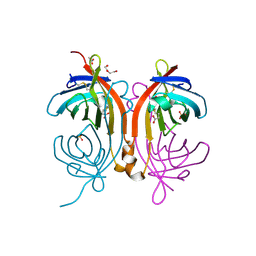
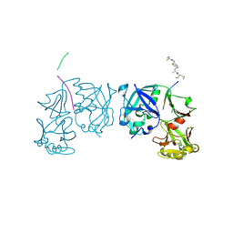
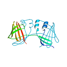
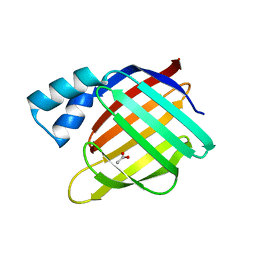
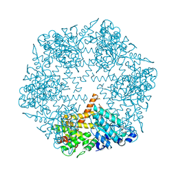
3KDS
DownloadVisualize
BU of 3kds by Molmil
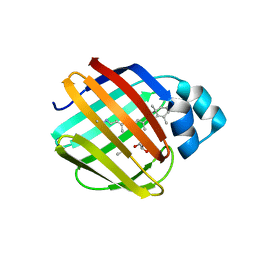
apo-FtsH crystal structure
分子名称: Cell division protein FtsH, N-{(2R)-2-[2-(hydroxyamino)-2-oxoethyl]-4-methylpentanoyl}-3-naphthalen-2-yl-L-alanyl-L-alaninamide, ZINC ION
著者Bieniossek, C, Niederhauser, B, Baumann, U.
登録日2009-10-23
公開日2009-12-01
最終更新日2023-11-01
実験手法X-RAY DIFFRACTION (2.601 Å)
主引用文献The crystal structure of apo-FtsH reveals domain movements necessary for substrate unfolding and translocation
Proc.Natl.Acad.Sci.USA, 106, 2009
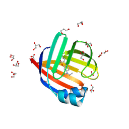
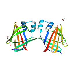
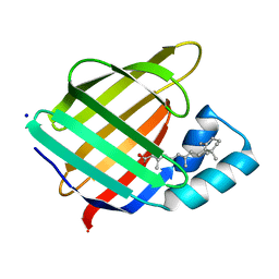
4BCS
DownloadVisualize
BU of 4bcs by Molmil
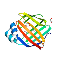
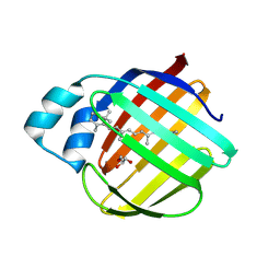
Crystal structure of an avidin mutant
分子名称: ACETATE ION, BIOTIN, CHIMERIC AVIDIN, ...
著者Airenne, T.T, Niederhauser, B, Hytonen, V.P, Kulomaa, M.S, Johnson, M.S.
登録日2012-10-03
公開日2013-10-16
最終更新日2023-12-20
実験手法X-RAY DIFFRACTION (1.8 Å)
主引用文献A Novel Chimeric Avidin with Increased Thermal Stability Using DNA Shuffling.
Plos One, 9, 2014
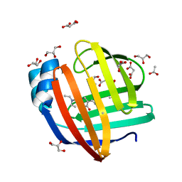
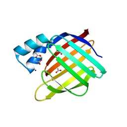
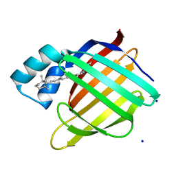
8D6H
DownloadVisualize
BU of 8d6h by Molmil
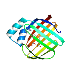
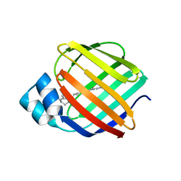
Q108K:K40L:T51C:T53A:R58L:Q38F:Q4F mutant of hCRBPII bound to synthetic fluorophore CM1V after UV irradiation
分子名称: (2E)-3-[7-(diethylamino)-2-oxo-2H-1-benzopyran-3-yl]prop-2-enal, bound form, ACETATE ION, ...
著者Bingham, C.R, Geiger, J.H, Borhan, B.
登録日2022-06-06
公開日2023-02-01
最終更新日2023-10-25
実験手法X-RAY DIFFRACTION (1.6 Å)
主引用文献Light controlled reversible Michael addition of cysteine: a new tool for dynamic site-specific labeling of proteins.
Analyst, 148, 2023
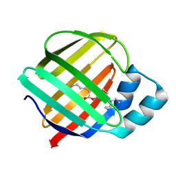
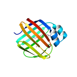
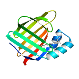
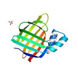
8D6N
DownloadVisualize
BU of 8d6n by Molmil
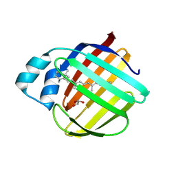
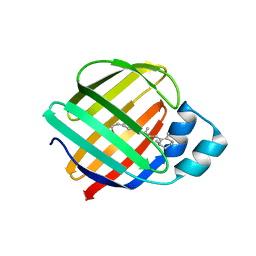
Q108K:K40L:T53A:R58L:Q38F:Q4F mutant of hCRBPII bound to synthetic fluorophore CM1V
分子名称: (2E)-3-[7-(diethylamino)-2-oxo-2H-1-benzopyran-3-yl]prop-2-enal, bound form, ACETATE ION, ...
著者Bingham, C.R, Geiger, J.H, Borhan, B.
登録日2022-06-06
公開日2023-02-01
最終更新日2023-10-25
実験手法X-RAY DIFFRACTION (1.42 Å)
主引用文献Light controlled reversible Michael addition of cysteine: a new tool for dynamic site-specific labeling of proteins.
Analyst, 148, 2023
8DB2
DownloadVisualize
BU of 8db2 by Molmil
Q108K:K40L:T51C:T53A:R58L:Q38F mutant of hCRBPII bound to synthetic fluorophore CM1V
分子名称: (2E)-3-[7-(diethylamino)-2-oxo-2H-1-benzopyran-3-yl]prop-2-enal, bound form, Retinol-binding protein 2
著者Bingham, C.R, Geiger, J.H, Borhan, B, Staples, R.
登録日2022-06-14
公開日2023-02-01
最終更新日2023-10-25
実験手法X-RAY DIFFRACTION (1.5 Å)
主引用文献Light controlled reversible Michael addition of cysteine: a new tool for dynamic site-specific labeling of proteins.
Analyst, 148, 2023
8D6L
DownloadVisualize
BU of 8d6l by Molmil
Q108K:K40L:T51C:T53A:R58L:Q38F:Q4F mutant of hCRBPII bound to synthetic fluorophore CM1V
分子名称: (2E)-3-[7-(diethylamino)-2-oxo-2H-1-benzopyran-3-yl]prop-2-enal, bound form, GLYCEROL, ...
著者Bingham, C.R, Geiger, J.H, Borhan, B.
登録日2022-06-06
公開日2023-02-15
最終更新日2023-10-25
実験手法X-RAY DIFFRACTION (1.69 Å)
主引用文献Light controlled reversible Michael addition of cysteine: a new tool for dynamic site-specific labeling of proteins.
Analyst, 148, 2023
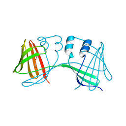
5DPQ
DownloadVisualize
BU of 5dpq by Molmil
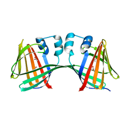
Crystal Structure of E72A mutant of domain swapped dimer Human Cellular Retinol Binding Protein
分子名称: ACETATE ION, Retinol-binding protein 2
著者Assar, Z, Nossoni, Z, Wang, W, Geiger, J.H, Borhan, B.
登録日2015-09-14
公開日2016-09-14
最終更新日2024-03-06
実験手法X-RAY DIFFRACTION (1.775 Å)
主引用文献Domain-Swapped Dimers of Intracellular Lipid-Binding Proteins: Evidence for Ordered Folding Intermediates.
Structure, 24, 2016
7LHJ
DownloadVisualize
BU of 7lhj by Molmil
Crystal structure of Q108K:K40L:T51V:T53C:R58W:T29L:Y19W:Q4A mutant of cellular retinol binding protein II complex with 15-cis-retinal
分子名称: ACETATE ION, GLYCEROL, RETINAL, ...
著者Ehyaei, N, Geiger, J.H, Borhan, B.
登録日2021-01-25
公開日2021-02-10
最終更新日2023-10-18
実験手法X-RAY DIFFRACTION (1.26 Å)
主引用文献Crystal structure of Q108K:K40L:T51V:T53C:R58W:T29L:Y19W:Q4A mutant of cellular retinol binding protein II complex with 15cis- retinal
To Be Published
7LHN
DownloadVisualize
BU of 7lhn by Molmil
Crystal structure of Q108K:K40L:T51V:T53C:R58W:T29L:Y19W:Q4A mutant of cellular retinol binding protein II complex with all-trans-retinal after exposure to visible light
分子名称: GLYCEROL, RETINAL, Retinol-binding protein 2
著者Ehyaei, N, Geiger, J.H, Borhan, B.
登録日2021-01-25
公開日2021-04-14
最終更新日2023-10-18
実験手法X-RAY DIFFRACTION (2.11 Å)
主引用文献Crystal structure of Q108K:K40L:T51V:T53C:R58W:T29L:Y19W:Q4A mutant of cellular retinol binding protein II complex with all-trans-retinal after exposure to visible light
To Be Published
7LHM
DownloadVisualize
BU of 7lhm by Molmil
Crystal structure of Q108K:K40L:T51V:T53C:R58W:T29L:Y19W:Q4A:Q38L mutant of cellular retinol binding protein II complex with all-trans-retinal
分子名称: ACETATE ION, GLYCEROL, RETINAL, ...
著者Ehyaei, N, Geiger, J.H, Borhan, B.
登録日2021-01-25
公開日2021-04-14
最終更新日2023-10-18
実験手法X-RAY DIFFRACTION (1.5 Å)
主引用文献Crystal structure of Q108K:K40L:T51V:T53C:R58W:T29L:Y19W:Q4A:Q38L mutant of cellular retinol binding protein II complex with all-trans-retinal
To Be Published
7LHO
DownloadVisualize
BU of 7lho by Molmil
Crystal structure of Q108K:K40L:T51V:T53C:R58W:T29L:Y19W:Q4A mutant of cellular retinol binding protein II complex with all-trans-retinal in the dark
分子名称: GLYCEROL, RETINAL, Retinol-binding protein 2
著者Ehyaei, N, Geiger, J.H, Borhan, B.
登録日2021-01-26
公開日2021-04-14
最終更新日2023-10-18
実験手法X-RAY DIFFRACTION (1.4 Å)
主引用文献Crystal structure of Q108K:K40L:T51V:T53C:R58W:T29L:Y19W:Q4A mutant of cellular retinol binding protein II complex with all-trans-retinal in the dark
To Be Published
4QYP
DownloadVisualize
BU of 4qyp by Molmil
The Crystal Structures of holo-wt human Cellular Retinol Binding protein II (hCRBPII) bound to Retinal
分子名称: ACETATE ION, RETINAL, Retinol-binding protein 2
著者Nossoni, Z, Assar, Z, Yapici, I, Nosrati, M, Wang, W, Berbasova, T, Vasileiou, C, Borhan, B, Geiger, H.
登録日2014-07-25
公開日2014-12-10
最終更新日2023-09-20
実験手法X-RAY DIFFRACTION (1.62 Å)
主引用文献Structures of holo wild-type human cellular retinol-binding protein II (hCRBPII) bound to retinol and retinal.
Acta Crystallogr.,Sect.D, 70, 2014
4QYN
DownloadVisualize
BU of 4qyn by Molmil
The Crystal Structures of holo-wt human Cellular Retinol Binding protein II (hCRBPII) bound to Retinol
分子名称: ACETATE ION, RETINOL, Retinol-binding protein 2
著者Nossoni, Z, Assar, Z, Yapici, I, Nosrati, M, Wang, W, Berbasova, T, Vasileiou, C, Borhan, B, Geiger, H.
登録日2014-07-24
公開日2014-12-31
最終更新日2024-02-28
実験手法X-RAY DIFFRACTION (1.19 Å)
主引用文献Structures of holo wild-type human cellular retinol-binding protein II (hCRBPII) bound to retinol and retinal.
Acta Crystallogr.,Sect.D, 70, 2014
1TZS
DownloadVisualize
BU of 1tzs by Molmil
Crystal Structure of an activation intermediate of Cathepsin E
分子名称: 23-mer peptide from PelB-IgG kappa light chain fusion protein, Cathepsin E, activation peptide from Cathepsin E
著者Ostermann, N, Gerhartz, B, Worpenberg, S, Trappe, J, Eder, J.
登録日2004-07-12
公開日2005-07-12
最終更新日2023-10-25
実験手法X-RAY DIFFRACTION (2.35 Å)
主引用文献Crystal structure of an activation intermediate of cathepsin e
J.Mol.Biol., 342, 2004
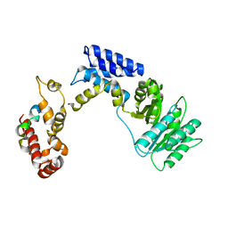
3KL4
DownloadVisualize
BU of 3kl4 by Molmil
Recognition of a signal peptide by the signal recognition particle
分子名称: Signal peptide of yeast dipeptidyl aminopeptidase B, Signal recognition 54 kDa protein
著者Janda, C.Y, Nagai, K, Li, J, Oubridge, C.
登録日2009-11-06
公開日2010-03-31
最終更新日2024-02-21
実験手法X-RAY DIFFRACTION (3.5 Å)
主引用文献Recognition of a signal peptide by the signal recognition particle.
Nature, 465, 2010
7LSQ
DownloadVisualize
BU of 7lsq by Molmil
The Crystal Structure of Q108K:K40E:T53A:R58W:Q38F:Q4F:Y19W Mutant of HCRBPII Bound with LizFluor Chromophore Showing Excited State Intermolecular Proton Transfer
分子名称: 4-{5-[(2E)-but-2-en-2-yl]thiophen-2-yl}-N,N-dimethylaniline, ACETATE ION, Retinol-binding protein 2
著者Ghanbarpour, A, Geiger, J.
登録日2021-02-18
公開日2022-01-12
最終更新日2023-10-18
実験手法X-RAY DIFFRACTION (1.67130542 Å)
主引用文献Design of Large Stokes Shift Fluorescent Proteins Based on Excited State Proton Transfer of an Engineered Photobase.
J.Am.Chem.Soc., 143, 2021
4ZH6
DownloadVisualize
BU of 4zh6 by Molmil
Crystal Structure of the Domain-Swapped Dimer Y60L mutant of Human Cellular Retinol Binding Protein II
分子名称: ACETATE ION, Retinol-binding protein 2
著者Assar, Z, Nossoni, Z, Geiger, J.H.
登録日2015-04-24
公開日2016-06-08
最終更新日2023-09-27
実験手法X-RAY DIFFRACTION (1.5497 Å)
主引用文献Domain-Swapped Dimers of Intracellular Lipid-Binding Proteins: Evidence for Ordered Folding Intermediates.
Structure, 24, 2016
4ZH9
DownloadVisualize
BU of 4zh9 by Molmil
Crystal Structure of the Domain-Swapped Dimer Wild-Type of Human Cellular Retinol Binding Protein II
分子名称: Retinol-binding protein 2
著者Assar, Z, Nossoni, Z, Geiger, J.H.
登録日2015-04-24
公開日2016-06-08
最終更新日2023-09-27
実験手法X-RAY DIFFRACTION (2.66 Å)
主引用文献Domain-Swapped Dimers of Intracellular Lipid-Binding Proteins: Evidence for Ordered Folding Intermediates.
Structure, 24, 2016
4ZR2
DownloadVisualize
BU of 4zr2 by Molmil
Crystal Structure of the Domain-Swapped Dimer K40L:Q108K:Y60W mutant of Human Cellular Retinol Binding Protein II
分子名称: ACETATE ION, RETINAL, Retinol-binding protein 2
著者Assar, Z, Nossoni, Z, Geiger, J.H.
登録日2015-05-11
公開日2016-06-08
最終更新日2023-09-27
実験手法X-RAY DIFFRACTION (1.8024 Å)
主引用文献Domain-Swapped Dimers of Intracellular Lipid-Binding Proteins: Evidence for Ordered Folding Intermediates.
Structure, 24, 2016
2FS6
DownloadVisualize
BU of 2fs6 by Molmil
Crystal Structure of Apo-Cellular Retinoic Acid Binding Protein Type II At 1.35 Angstroms Resolution
分子名称: ACETATE ION, CHLORIDE ION, Cellular retinoic acid-binding protein 2, ...
著者Vaezeslami, S, Geiger, J.H.
登録日2006-01-21
公開日2006-09-19
最終更新日2024-02-14
実験手法X-RAY DIFFRACTION (1.35 Å)
主引用文献The structure of Apo-wild-type cellular retinoic acid binding protein II at 1.4 A and its relationship to ligand binding and nuclear translocation.
J.Mol.Biol., 363, 2006
2FR3
DownloadVisualize
BU of 2fr3 by Molmil
Crystal Structure of Cellular Retinoic Acid Binding Protein Type II in Complex with All-Trans-Retinoic Acid at 1.48 Angstroms Resolution
分子名称: ACETATE ION, Cellular retinoic acid binding protein 2, RETINOIC ACID
著者Vaezeslami, S, Geiger, J.H.
登録日2006-01-18
公開日2006-09-19
最終更新日2024-02-14
実験手法X-RAY DIFFRACTION (1.48 Å)
主引用文献The structure of Apo-wild-type cellular retinoic acid binding protein II at 1.4 A and its relationship to ligand binding and nuclear translocation.
J.Mol.Biol., 363, 2006
2FS7
DownloadVisualize
BU of 2fs7 by Molmil
Crystal Structure of Apo-Cellular Retinoic Acid Binding Protein Type II At 1.55 Angstroms Resolution
分子名称: ACETATE ION, CHLORIDE ION, Cellular retinoic acid-binding protein 2
著者Vaezeslami, S, Geiger, J.H.
登録日2006-01-21
公開日2006-09-19
最終更新日2024-02-14
実験手法X-RAY DIFFRACTION (1.55 Å)
主引用文献The structure of Apo-wild-type cellular retinoic acid binding protein II at 1.4 A and its relationship to ligand binding and nuclear translocation.
J.Mol.Biol., 363, 2006
2G78
DownloadVisualize
BU of 2g78 by Molmil
Crystal Structure of the R132K:Y134F Mutant of Cellular Retinoic Acid Binding Protein Type II in Complex with All-Trans-Retinoic Acid at 1.70 Angstroms Resolution
分子名称: Cellular retinoic acid-binding protein 2, RETINOIC ACID, SODIUM ION
著者Vaezeslami, S, Geiger, J.H.
登録日2006-02-27
公開日2007-04-17
最終更新日2024-02-14
実験手法X-RAY DIFFRACTION (1.7 Å)
主引用文献Protein design: reengineering cellular retinoic acid binding protein II into a rhodopsin protein mimic.
J.Am.Chem.Soc., 129, 2007
2G7B
DownloadVisualize
BU of 2g7b by Molmil
Crystal Structure of the R132K:R111L:L121E mutant of Cellular Retinoic Acid Binding Protein Type II In Complex With All-Trans-Retinal At 1.18 Angstroms Resolution
分子名称: ALL-TRANS AXEROPHTHENE, Cellular retinoic acid-binding protein 2, SODIUM ION
著者Vaezeslami, S, Geiger, J.H.
登録日2006-02-27
公開日2007-04-17
最終更新日2021-10-20
実験手法X-RAY DIFFRACTION (1.18 Å)
主引用文献Protein design: reengineering cellular retinoic acid binding protein II into a rhodopsin protein mimic.
J.Am.Chem.Soc., 129, 2007
2G79
DownloadVisualize
BU of 2g79 by Molmil
Crystal Structure of the R132K:Y134F Mutant of Cellular Retinoic Acid Binding Protein Type II in Complex with All-Trans-Retinal at 1.69 Angstroms Resolution
分子名称: Cellular retinoic acid-binding protein 2, RETINAL, SODIUM ION, ...
著者Vaezeslami, S, Geiger, J.H.
登録日2006-02-27
公開日2007-04-17
最終更新日2024-02-14
実験手法X-RAY DIFFRACTION (1.69 Å)
主引用文献Protein design: reengineering cellular retinoic acid binding protein II into a rhodopsin protein mimic.
J.Am.Chem.Soc., 129, 2007

222415

件を2024-07-10に公開中

PDB statisticsPDBj update infoContact PDBjnumon