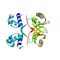
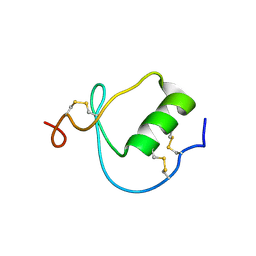
1HO2
 
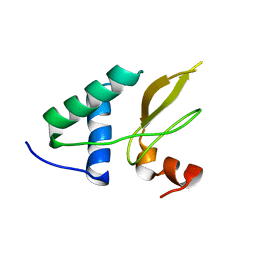
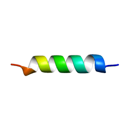 | | NMR STRUCTURE OF THE POTASSIUM CHANNEL FRAGMENT L45 IN MICELLES | | 分子名称: | VOLTAGE-GATED POTASSIUM CHANNEL PROTEIN | | 著者 | Ohlenschlager, O, Hojo, H, Ramachandran, R, Gorlach, M, Haris, P.I. | | 登録日 | 2000-12-08 | | 公開日 | 2002-06-05 | | 最終更新日 | 2024-05-22 | | 実験手法 | SOLUTION NMR | | 主引用文献 | Three-dimensional structure of the S4-S5 segment of the Shaker potassium channel.
Biophys.J., 82, 2002
|
|
1UUU
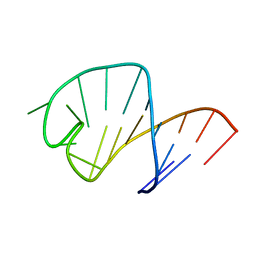 | | STRUCTURE OF AN RNA HAIRPIN LOOP WITH A 5'-CGUUUCG-3' LOOP MOTIF BY HETERONUCLEAR NMR SPECTROSCOPY AND DISTANCE GEOMETRY, 15 STRUCTURES | | 分子名称: | RNA (5'-R(*GP*GP*CP*GP*UP*AP*CP*GP*UP*UP*UP*CP*GP*UP*AP*CP*GP*CP*C)-3') | | 著者 | Sich, C, Ohlenschlager, O, Ramachandran, R, Gorlach, M, Brown, L.R. | | 登録日 | 1997-08-12 | | 公開日 | 1998-02-25 | | 最終更新日 | 2024-05-22 | | 実験手法 | SOLUTION NMR | | 主引用文献 | Structure of an RNA hairpin loop with a 5'-CGUUUCG-3' loop motif by heteronuclear NMR spectroscopy and distance geometry.
Biochemistry, 36, 1997
|
|
1HO7
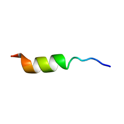 | | NMR STRUCTURE OF THE POTASSIUM CHANNEL FRAGMENT L45 IN TFE | | 分子名称: | VOLTAGE-GATED POTASSIUM CHANNEL PROTEIN | | 著者 | Ohlenschlager, O, Hojo, H, Ramachandran, R, Gorlach, M, Haris, P.I. | | 登録日 | 2000-12-10 | | 公開日 | 2002-06-05 | | 最終更新日 | 2024-05-22 | | 実験手法 | SOLUTION NMR | | 主引用文献 | Three-dimensional structure of the S4-S5 segment of the Shaker potassium channel.
Biophys.J., 82, 2002
|
|
1ZAU
 | |
2VBX
 | |
2VC1
 | |
2VBZ
 | |
2VBY
 | |
2VC0
 | |
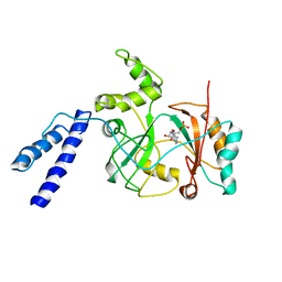
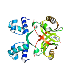
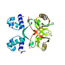
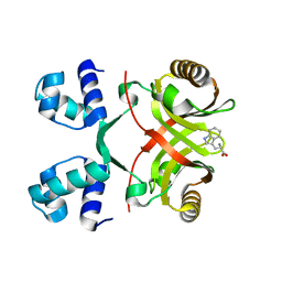
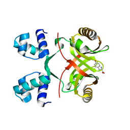
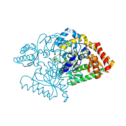
2VOJ
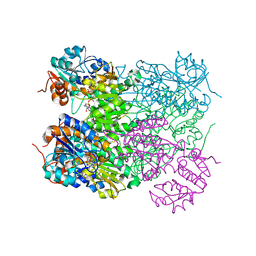 | | Ternary complex of M. tuberculosis Rv2780 with NAD and pyruvate | | 分子名称: | (2S)-2-HYDROXYPROPANOIC ACID, ALANINE DEHYDROGENASE, NICOTINAMIDE-ADENINE-DINUCLEOTIDE | | 著者 | Tripathi, S.M, Ramachandran, R. | | 登録日 | 2008-02-18 | | 公開日 | 2008-03-04 | | 最終更新日 | 2024-05-08 | | 実験手法 | X-RAY DIFFRACTION (2.6 Å) | | 主引用文献 | Crystal Structures of the Mycobacterium Tuberculosis Secretory Antigen Alanine Dehydrogenase (Rv2780) in Apo and Ternary Complex Forms Captures "Open" and "Closed" Enzyme Conformations.
Proteins: Struct., Funct., Bioinf., 72, 2008
|
|
2W25
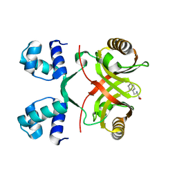
 | | Crystal structure of Glu104Ala mutant | | 分子名称: | PROBABLE TRANSCRIPTIONAL REGULATORY PROTEIN | | 著者 | Shrivastava, T, RAmachandran, R. | | 登録日 | 2008-10-24 | | 公開日 | 2009-11-17 | | 最終更新日 | 2023-12-13 | | 実験手法 | X-RAY DIFFRACTION (2.15 Å) | | 主引用文献 | Ligand-Induced Structural Transitions, Mutational Analysis, and 'Open' Quaternary Structure of the M. Tuberculosis Feast/Famine Regulatory Protein (Rv3291C).
J.Mol.Biol., 392, 2009
|
|
2W24
 | | M. tuberculosis Rv3291c complexed to Lysine | | 分子名称: | LYSINE, PROBABLE TRANSCRIPTIONAL REGULATORY PROTEIN | | 著者 | Shrivastava, T, Ramachandran, R. | | 登録日 | 2008-10-24 | | 公開日 | 2009-11-17 | | 最終更新日 | 2023-12-13 | | 実験手法 | X-RAY DIFFRACTION (2.5 Å) | | 主引用文献 | Ligand-Induced Structural Transitions, Mutational Analysis, and 'Open' Quaternary Structure of the M. Tuberculosis Feast/Famine Regulatory Protein (Rv3291C).
J.Mol.Biol., 392, 2009
|
|
2VBW
 | |
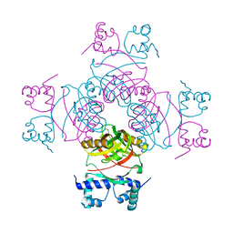
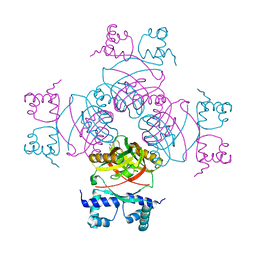
2XI1
 | | Crystal structure of the HIV-1 Nef sequenced from a patient's sample | | 分子名称: | NEF | | 著者 | Yadav, G.P, Singh, P, Gupta, S, Tripathi, A.K, Tripathi, R.K, Ramachandran, R. | | 登録日 | 2010-06-25 | | 公開日 | 2011-08-10 | | 最終更新日 | 2024-05-08 | | 実験手法 | X-RAY DIFFRACTION (3.5 Å) | | 主引用文献 | A Novel Dimer-Tetramer Transition Captured by the Crystal Structure of the HIV-1 Nef.
Plos One, 6, 2011
|
|
2VU5
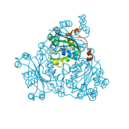 | | Crystal structure of Pndk from Bacillus anthracis | | 分子名称: | NUCLEOSIDE DIPHOSPHATE KINASE | | 著者 | Misra, G, Aggarwal, A, Dube, D, Zaman, M.S, Singh, Y, Ramachandran, R. | | 登録日 | 2008-05-21 | | 公開日 | 2009-03-10 | | 最終更新日 | 2024-05-08 | | 実験手法 | X-RAY DIFFRACTION (2 Å) | | 主引用文献 | Crystal Structure of the Bacillus Anthracis Nucleoside Diphosphate Kinase and its Characterization Reveals an Enzyme Adapted to Perform Under Stress Conditions.
Proteins, 76, 2009
|
|
2VOE
 | |
1KMA
 | | NMR Structure of the Domain-I of the Kazal-type Thrombin Inhibitor Dipetalin | | 分子名称: | DIPETALIN | | 著者 | Schlott, B, Wohnert, J, Icke, C, Hartmann, M, Ramachandran, R, Guhrs, K.-H, Glusa, E, Flemming, J, Gorlach, M, Grosse, F, Ohlenschlager, O. | | 登録日 | 2001-12-14 | | 公開日 | 2002-05-15 | | 最終更新日 | 2022-02-23 | | 実験手法 | SOLUTION NMR | | 主引用文献 | Interaction of Kazal-type inhibitor domains with serine proteinases: biochemical and structural studies.
J.Mol.Biol., 318, 2002
|
|
2CIN
 | |
2CJH
 | |
2CJD
 | |
2CJG
 | |
2IVM
 | |
2JJF
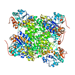
 | | N328A mutant of M. tuberculosis Rv3290c | | 分子名称: | L-LYSINE EPSILON AMINOTRANSFERASE | | 著者 | tripathi, S.M, Ramachandran, R. | | 登録日 | 2008-04-04 | | 公開日 | 2009-06-30 | | 最終更新日 | 2024-05-08 | | 実験手法 | X-RAY DIFFRACTION (1.95 Å) | | 主引用文献 | Mutational Analysis of Mycobacterium Tuberculosis Lysine Epsilon-Aminotransferase and Inhibitor Co-Crystal Structures, Reveals Distinct Binding Modes.
Biochem.Biophys.Res.Commun., 463, 2015
|
|
2JJH
 | | E243 mutant of M. tuberculosis Rv3290C | | 分子名称: | 2-OXOGLUTARIC ACID, L-LYSINE EPSILON AMINOTRANSFERASE, PYRIDOXAL-5'-PHOSPHATE | | 著者 | Tripathi, S.M, Ramachandran, R. | | 登録日 | 2008-04-04 | | 公開日 | 2009-06-30 | | 最終更新日 | 2020-01-15 | | 実験手法 | X-RAY DIFFRACTION (2.7 Å) | | 主引用文献 | Mutational Analysis of Mycobacterium Tuberculosis Lysine Epsilon-Aminotransferase and Inhibitor Co-Crystal Structures, Reveals Distinct Binding Modes.
Biochem.Biophys.Res.Commun., 463, 2015
|
|
2JJE
 | |


