6DFV
 
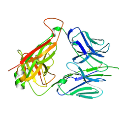 | | Mouse diabetogenic TCR 8F10 | | 分子名称: | 1,2-ETHANEDIOL, TCR alpha chain, TCR beta chain | | 著者 | Wang, Y, Dai, S. | | 登録日 | 2018-05-15 | | 公開日 | 2019-04-17 | | 最終更新日 | 2024-11-13 | | 実験手法 | X-RAY DIFFRACTION (1.71 Å) | | 主引用文献 | How C-terminal additions to insulin B-chain fragments create superagonists for T cells in mouse and human type 1 diabetes.
Sci Immunol, 4, 2019
|
|
6DFS
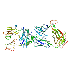 | | mouse TCR I.29 in complex with IAg7-p8E9E6ss | | 分子名称: | 2-acetamido-2-deoxy-beta-D-glucopyranose, H-2 class II histocompatibility antigen, A-D alpha chain, ... | | 著者 | Wang, Y, Dai, S. | | 登録日 | 2018-05-15 | | 公開日 | 2019-04-17 | | 最終更新日 | 2024-10-09 | | 実験手法 | X-RAY DIFFRACTION (3.1 Å) | | 主引用文献 | How C-terminal additions to insulin B-chain fragments create superagonists for T cells in mouse and human type 1 diabetes.
Sci Immunol, 4, 2019
|
|
6DFW
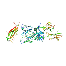 | | TCR 8F10 in complex with IAg7-p8G9E | | 分子名称: | 8F10 alpha chain, 8F10 beta chain, H-2 class II histocompatibility antigen, ... | | 著者 | Wang, Y, Dai, S. | | 登録日 | 2018-05-15 | | 公開日 | 2019-04-17 | | 最終更新日 | 2024-11-06 | | 実験手法 | X-RAY DIFFRACTION (3.2 Å) | | 主引用文献 | How C-terminal additions to insulin B-chain fragments create superagonists for T cells in mouse and human type 1 diabetes.
Sci Immunol, 4, 2019
|
|
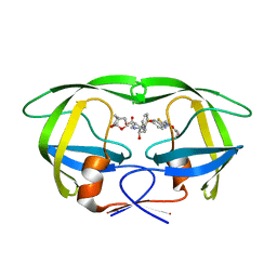
1IXD
 | | Solution structure of the CAP-GLY domain from human cylindromatosis tomour-suppressor CYLD | | 分子名称: | Cylindromatosis tumour-suppressor CYLD | | 著者 | Saito, K, Koshiba, S, Kigawa, T, Yokoyama, S, RIKEN Structural Genomics/Proteomics Initiative (RSGI) | | 登録日 | 2002-06-19 | | 公開日 | 2002-12-19 | | 最終更新日 | 2023-12-27 | | 実験手法 | SOLUTION NMR | | 主引用文献 | The CAP-Gly domain of CYLD associates with the proline-rich sequence in NEMO/IKKgamma
STRUCTURE, 12, 2004
|
|
6MK9
 | | X-ray crystal structure of darunavir-resistant-P51 HIV-1 protease in complex with GRL-121 | | 分子名称: | (3S,3aR,5R,7aS,8S)-hexahydro-4H-3,5-methanofuro[2,3-b]pyran-8-yl {(2S,3R)-4-[{[2-(cyclopropylamino)-1,3-benzothiazol-6-yl]sulfonyl}(2-methylpropyl)amino]-3-hydroxy-1-phenylbutan-2-yl}carbamate, Protease | | 著者 | Yedidi, R.S, Hayashi, H, Das, D, Mitsuya, H. | | 登録日 | 2018-09-25 | | 公開日 | 2019-10-02 | | 最終更新日 | 2024-12-25 | | 実験手法 | X-RAY DIFFRACTION (1.7 Å) | | 主引用文献 | X-ray crystal structure of darunavir-resistant-P51 HIV-1 protease in complex with GRL-121
To Be Published
|
|
6MKL
 | | X-ray crystal structure of darunavir-resistant-P51 HIV-1 protease in complex with GRL-142 | | 分子名称: | (3S,3aR,5R,7aS,8S)-hexahydro-4H-3,5-methanofuro[2,3-b]pyran-8-yl [(2S,3R)-4-[{[2-(cyclopropylamino)-1,3-benzothiazol-6-yl]sulfonyl}(2-methylpropyl)amino]-1-(3,5-difluorophenyl)-3-hydroxybutan-2-yl]carbamate, Protease | | 著者 | Yedidi, R.S, Hayashi, H, Das, D, Mitsuya, H. | | 登録日 | 2018-09-25 | | 公開日 | 2019-10-02 | | 最終更新日 | 2023-10-11 | | 実験手法 | X-RAY DIFFRACTION (1.7 Å) | | 主引用文献 | Structure-function analysis of darunavir-resistant-P51 HIV-1 protease in complex with GRL-142.
To Be Published
|
|


