6UN7
 
 | |
6UMY
 | |
6UN5
 | |
6UN6
 | |
6UN2
 | |
6UN4
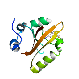
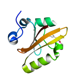
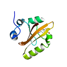
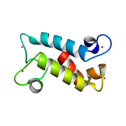
 | | Crystal structure of rsEGFP2, Y67(3-ClY), Y107(3-ClY) | | 分子名称: | Green fluorescent protein, SULFATE ION | | 著者 | Lin, C.-Y, Romei, M.G, Boxer, S.G, Chang, J. | | 登録日 | 2019-10-10 | | 公開日 | 2020-07-15 | | 最終更新日 | 2023-11-15 | | 実験手法 | X-RAY DIFFRACTION (1.499 Å) | | 主引用文献 | Structural and spectroscopic characterization of photoactive yellow protein and photoswitchable fluorescent protein constructs containing heavy atoms.
J Photochem Photobiol A Chem, 401, 2020
|
|
6UN0
 | |
8FNR
 | |
8FNS
 | |
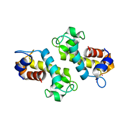
8DQ2
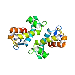 | | X-ray crystal structure of Hansschlegelia quercus lanmodulin (LanM) with lanthanum (III) bound at pH 7 | | 分子名称: | CITRIC ACID, EF-hand domain-containing protein, LANTHANUM (III) ION, ... | | 著者 | Jung, J.J, Lin, C.-Y, Boal, A.K. | | 登録日 | 2022-07-18 | | 公開日 | 2023-06-07 | | 最終更新日 | 2024-05-22 | | 実験手法 | X-RAY DIFFRACTION (1.8 Å) | | 主引用文献 | Enhanced rare-earth separation with a metal-sensitive lanmodulin dimer.
Nature, 618, 2023
|
|
4CLC
 | |


