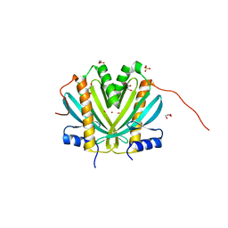
2OXJ
 
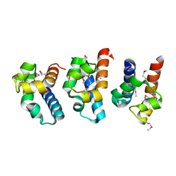
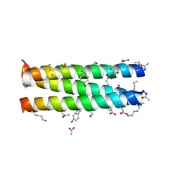 | | Helix Bundle Quaternary Structure from alpha/beta-Peptide Foldamers: GCN4-p1 with beta-residues at b and f heptad positions. | | 分子名称: | ACETATE ION, hybrid alpha/beta peptide based on the GCN4-p1 sequence; heptad positions b and f substituted with beta-amino acids | | 著者 | Horne, W.S, Price, J.L, Keck, J.L, Gellman, S.H. | | 登録日 | 2007-02-20 | | 公開日 | 2007-03-27 | | 最終更新日 | 2024-07-10 | | 実験手法 | X-RAY DIFFRACTION (2 Å) | | 主引用文献 | Helix Bundle Quaternary Structure from alpha/beta-Peptide Foldamers.
J.Am.Chem.Soc., 129, 2007
|
|
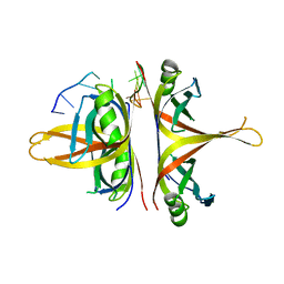
2OXK
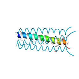 | | Helix Bundle Quaternary Structure from alpha/beta-Peptide Foldamers: GCN4-pLI with beta-residues at b and f heptad positions. | | 分子名称: | FORMIC ACID, hybrid alpha/beta peptide based on the GCN4-pLI sequence; heptad positions b and f substituted with beta-amino acids | | 著者 | Horne, W.S, Price, J.L, Keck, J.L, Gellman, S.H. | | 登録日 | 2007-02-20 | | 公開日 | 2007-03-27 | | 最終更新日 | 2024-07-10 | | 実験手法 | X-RAY DIFFRACTION (2 Å) | | 主引用文献 | Helix Bundle Quaternary Structure from alpha/beta-Peptide Foldamers.
J.Am.Chem.Soc., 129, 2007
|
|
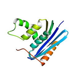
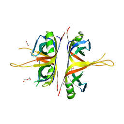
3NCT
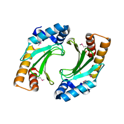 | | X-ray crystal structure of the bacterial conjugation factor PsiB, a negative regulator of reca | | 分子名称: | Protein psiB | | 著者 | Petrova, V, Satyshur, K.A, George, N.P, McCaslin, D, Cox, M.M, Keck, J.L. | | 登録日 | 2010-06-05 | | 公開日 | 2010-07-21 | | 最終更新日 | 2011-07-13 | | 実験手法 | X-RAY DIFFRACTION (2.2 Å) | | 主引用文献 | X-ray crystal structure of the bacterial conjugation factor PsiB, a negative regulator of RecA.
J.Biol.Chem., 285, 2010
|
|
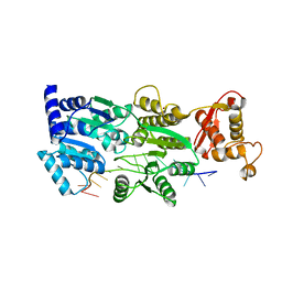
3MXN
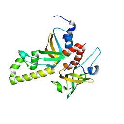 | | Crystal structure of the RMI core complex | | 分子名称: | BENZAMIDINE, RecQ-mediated genome instability protein 1, RecQ-mediated genome instability protein 2 | | 著者 | Hoadley, K.A, Xu, D, Xue, Y, Satyshur, K.A, Wang, W, Keck, J.L. | | 登録日 | 2010-05-07 | | 公開日 | 2010-09-15 | | 最終更新日 | 2024-02-21 | | 実験手法 | X-RAY DIFFRACTION (1.55 Å) | | 主引用文献 | Structure and cellular roles of the RMI core complex from the bloom syndrome dissolvasome.
Structure, 18, 2010
|
|
3IGM
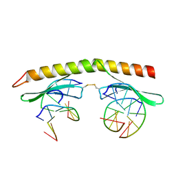 | | A 2.2A crystal structure of the AP2 domain of PF14_0633 from P. falciparum, bound as a domain-swapped dimer to its cognate DNA | | 分子名称: | 5'-D(*TP*GP*CP*AP*TP*GP*CP*A)-3', PF14_0633 protein | | 著者 | Lindner, S.E, De Silva, E, Keck, J.L, Llinas, M. | | 登録日 | 2009-07-28 | | 公開日 | 2009-11-10 | | 最終更新日 | 2017-11-01 | | 実験手法 | X-RAY DIFFRACTION (2.2 Å) | | 主引用文献 | Structural Determinants of DNA Binding by a P. falciparum ApiAP2 Transcriptional Regulator.
J.Mol.Biol., 395, 2010
|
|
1SE8
 | | Structure of single-stranded DNA-binding protein (SSB) from D. radiodurans | | 分子名称: | Single-strand binding protein | | 著者 | Bernstein, D.A, Eggington, J.M, Killoran, M.P, Misic, A.M, Cox, M.M, Keck, J.L. | | 登録日 | 2004-02-16 | | 公開日 | 2004-06-15 | | 最終更新日 | 2011-07-13 | | 実験手法 | X-RAY DIFFRACTION (1.8 Å) | | 主引用文献 | Crystal structure of the Deinococcus radiodurans single-stranded DNA-binding protein suggests a mechanism for coping with DNA damage.
Proc.Natl.Acad.Sci.USA, 101, 2004
|
|
3SXU
 | |
3PVS
 | | Structure and biochemical activities of Escherichia coli MgsA | | 分子名称: | PHOSPHATE ION, Replication-associated recombination protein A | | 著者 | Page, A.N, George, N.P, Marceau, A.H, Cox, M.M, Keck, J.L. | | 登録日 | 2010-12-07 | | 公開日 | 2011-02-02 | | 最終更新日 | 2024-02-21 | | 実験手法 | X-RAY DIFFRACTION (2.5 Å) | | 主引用文献 | Structure and Biochemical Activities of Escherichia coli MgsA.
J.Biol.Chem., 286, 2011
|
|
1WUD
 | | E. coli RecQ HRDC domain | | 分子名称: | ATP-dependent DNA helicase recQ | | 著者 | Bernstein, D.A, Keck, J.L. | | 登録日 | 2004-12-04 | | 公開日 | 2005-08-09 | | 最終更新日 | 2011-07-13 | | 実験手法 | X-RAY DIFFRACTION (2.2 Å) | | 主引用文献 | Conferring Substrate Specificity to DNA Helicases: Role of the RecQ HRDC Domain
STRUCTURE, 13, 2005
|
|
1F21
 | |
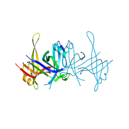
1YO8
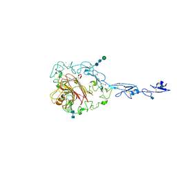 | | Structure of the C-terminal domain of human thrombospondin-2 | | 分子名称: | 2-acetamido-2-deoxy-beta-D-glucopyranose, 2-acetamido-2-deoxy-beta-D-glucopyranose-(1-4)-2-acetamido-2-deoxy-beta-D-glucopyranose, CALCIUM ION, ... | | 著者 | Carlson, C.B, Bernstein, D.A, Annis, D.S, Misenheimer, T.M, Hannah, B.A, Mosher, D.F, Keck, J.L. | | 登録日 | 2005-01-26 | | 公開日 | 2005-09-27 | | 最終更新日 | 2020-07-29 | | 実験手法 | X-RAY DIFFRACTION (2.6 Å) | | 主引用文献 | Structure of the calcium-rich signature domain of human thrombospondin-2
Nat.Struct.Mol.Biol., 12, 2005
|
|
1Z1A
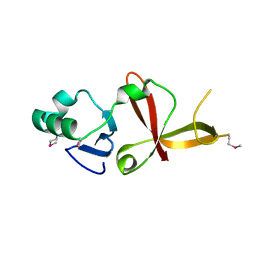 | |
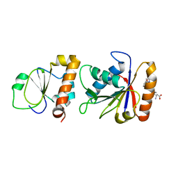
1ZHI
 | | Complex of the S. cerevisiae Orc1 and Sir1 interacting domains | | 分子名称: | Origin recognition complex subunit 1, Regulatory protein SIR1 | | 著者 | Hou, Z, Bernstein, D.A, Fox, C.A, Keck, J.L. | | 登録日 | 2005-04-25 | | 公開日 | 2005-06-07 | | 最終更新日 | 2024-02-14 | | 実験手法 | X-RAY DIFFRACTION (2.7 Å) | | 主引用文献 | Structural basis of the Sir1-origin recognition complex interaction in transcriptional silencing.
Proc.Natl.Acad.Sci.Usa, 102, 2005
|
|
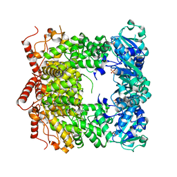
3C9D
 | | Crystal structure of Vps75 | | 分子名称: | Vacuolar protein sorting-associated protein 75 | | 著者 | Berndsen, C.E, Tsubota, T, Lindner, S.E, Lee, S, Holton, J.M, Kaufman, P.D, Keck, J.L, Denu, J.M. | | 登録日 | 2008-02-15 | | 公開日 | 2008-08-12 | | 最終更新日 | 2024-02-21 | | 実験手法 | X-RAY DIFFRACTION (2 Å) | | 主引用文献 | Molecular functions of the histone acetyltransferase chaperone complex Rtt109-Vps75
Nat.Struct.Mol.Biol., 15, 2008
|
|
3C94
 | | ExoI/SSB-Ct complex | | 分子名称: | Exodeoxyribonuclease I, MAGNESIUM ION, Single-stranded DNA-binding C-terminal tail peptide | | 著者 | Lu, D, Keck, J.L. | | 登録日 | 2008-02-15 | | 公開日 | 2008-07-08 | | 最終更新日 | 2023-08-30 | | 実験手法 | X-RAY DIFFRACTION (2.7 Å) | | 主引用文献 | Structural basis of Escherichia coli single-stranded DNA-binding protein stimulation of exonuclease I.
Proc.Natl.Acad.Sci.USA, 105, 2008
|
|
4DAY
 | |
2PNH
 | |
4H5B
 | | Crystal Structure of DR_1245 from Deinococcus radiodurans | | 分子名称: | BROMIDE ION, DR_1245 protein, GLYCEROL, ... | | 著者 | Norais, C, Servant, P, Bouthier-de-la-Tour, C, Coureux, P.D, Ithurbide, S, Vannier, F, Guerin, P, Dulberger, C.L, Satyshur, K.A, Keck, J.L, Armengaud, J, Cox, M.M, Sommer, S. | | 登録日 | 2012-09-18 | | 公開日 | 2013-01-30 | | 最終更新日 | 2024-02-28 | | 実験手法 | X-RAY DIFFRACTION (2 Å) | | 主引用文献 | The Deinococcus radiodurans DR1245 Protein, a DdrB Partner Homologous to YbjN Proteins and Reminiscent of Type III Secretion System Chaperones.
Plos One, 8, 2013
|
|
6BHX
 | | B. subtilis SsbA with DNA | | 分子名称: | DNA (5'-D(P*TP*TP*TP*TP*TP*TP*TP*TP*TP*TP*T)-3'), Single-stranded DNA-binding protein A | | 著者 | Dubiel, K.D, Myers, A.R, Satyshur, K.A, Keck, J.L. | | 登録日 | 2017-10-31 | | 公開日 | 2018-12-19 | | 最終更新日 | 2024-03-13 | | 実験手法 | X-RAY DIFFRACTION (2.936 Å) | | 主引用文献 | Structural Mechanisms of Cooperative DNA Binding by Bacterial Single-Stranded DNA-Binding Proteins.
J. Mol. Biol., 431, 2019
|
|
6BHW
 | | B. subtilis SsbA | | 分子名称: | 1,2-ETHANEDIOL, DI(HYDROXYETHYL)ETHER, Single-stranded DNA-binding protein A | | 著者 | Dubiel, K.D, Myers, A.R, Satyshur, K.A, Keck, J.L. | | 登録日 | 2017-10-31 | | 公開日 | 2018-12-19 | | 最終更新日 | 2023-10-04 | | 実験手法 | X-RAY DIFFRACTION (2.208 Å) | | 主引用文献 | Structural Mechanisms of Cooperative DNA Binding by Bacterial Single-Stranded DNA-Binding Proteins.
J. Mol. Biol., 431, 2019
|
|
6CRM
 | |
6D9Q
 | | The sulfate-bound crystal structure of HPRT (hypoxanthine phosphoribosyltransferase) | | 分子名称: | GLYCEROL, Hypoxanthine phosphoribosyltransferase, SULFATE ION | | 著者 | Satyshur, K.A, Dubiel, K, Anderson, B, Wolak, C, Keck, J.L. | | 登録日 | 2018-04-30 | | 公開日 | 2019-05-01 | | 最終更新日 | 2023-10-04 | | 実験手法 | X-RAY DIFFRACTION (2.056 Å) | | 主引用文献 | Evolution of (p)ppGpp-HPRT regulation through diversification of an allosteric oligomeric interaction.
Elife, 8, 2019
|
|
6DCR
 | |
4NL4
 | |
6DGD
 | | PriA helicase bound to dsDNA of a DNA replication fork | | 分子名称: | DNA (5'-D(P*AP*GP*CP*AP*CP*GP*CP*CP*GP*AP*CP*T)-3'), DNA (5'-D(P*GP*AP*GP*CP*AP*CP*GP*CP*CP*GP*AP*CP*T)-3'), DNA (5'-D(P*GP*TP*CP*GP*GP*CP*GP*TP*GP*CP*TP*C)-3'), ... | | 著者 | Satyshur, K.A, Windgassen, T.A, Keck, J.L. | | 登録日 | 2018-05-17 | | 公開日 | 2018-09-05 | | 最終更新日 | 2023-10-11 | | 実験手法 | X-RAY DIFFRACTION (2.823 Å) | | 主引用文献 | Structure-specific DNA replication-fork recognition directs helicase and replication restart activities of the PriA helicase.
Proc. Natl. Acad. Sci. U.S.A., 115, 2018
|
|


