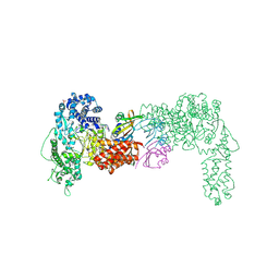
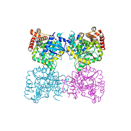
9H6C
 
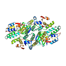
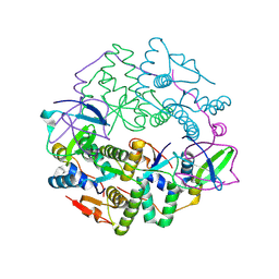 | | Crystal structure of the E. coli F-plasmid VapBC toxin-antitoxin complex (VapB T3N) | | 分子名称: | Antitoxin, tRNA(fMet)-specific endonuclease VapC | | 著者 | Hollingshead, S, McVicker, G, Nielsen, M.R, Zhang, Y, Pilla, G, Jones, R.A, Thomas, J.C, Johansen, S.E.H, Exley, R.M, Brodersen, D.E, Tang, C.M. | | 登録日 | 2024-10-24 | | 公開日 | 2024-12-18 | | 最終更新日 | 2025-02-12 | | 実験手法 | X-RAY DIFFRACTION (2.65 Å) | | 主引用文献 | Shared mechanisms of enhanced plasmid maintenance and antibiotic tolerance mediated by the VapBC toxin:antitoxin system.
Mbio, 16, 2025
|
|
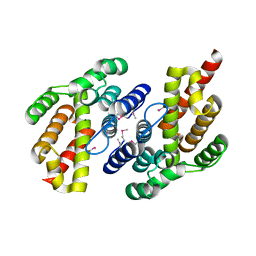
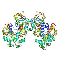
9H6D
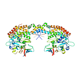
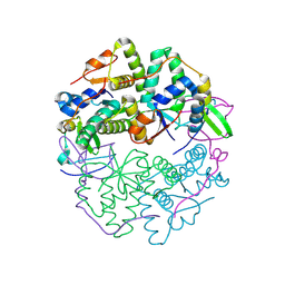 | | Crystal structure of the E. coli F-plasmid VapBC toxin-antitoxin complex (VapB V5E) | | 分子名称: | Antitoxin, tRNA(fMet)-specific endonuclease VapC | | 著者 | Hollingshead, S, McVicker, G, Nielsen, M.R, Zhang, Y, Pilla, G, Jones, R.A, Thomas, J.C, Johansen, S.E.H, Exley, R.M, Brodersen, D.E, Tang, C.M. | | 登録日 | 2024-10-24 | | 公開日 | 2024-12-18 | | 最終更新日 | 2025-02-12 | | 実験手法 | X-RAY DIFFRACTION (3.15 Å) | | 主引用文献 | Shared mechanisms of enhanced plasmid maintenance and antibiotic tolerance mediated by the VapBC toxin:antitoxin system.
Mbio, 16, 2025
|
|
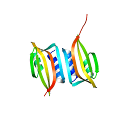
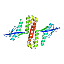
9H6B
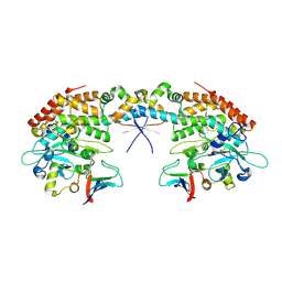
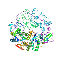 | | Crystal structure of the E. coli F-plasmid VapBC toxin-antitoxin complex (VapB T3N, A13P, L16R) | | 分子名称: | Antitoxin, tRNA(fMet)-specific endonuclease VapC | | 著者 | Hollingshead, S, McVicker, G, Nielsen, M.R, Zhang, Y, Pilla, G, Jones, R.A, Thomas, J.C, Johansen, S.E.H, Exley, R.M, Brodersen, D.E, Tang, C.M. | | 登録日 | 2024-10-24 | | 公開日 | 2024-12-18 | | 最終更新日 | 2025-02-12 | | 実験手法 | X-RAY DIFFRACTION (2.8 Å) | | 主引用文献 | Shared mechanisms of enhanced plasmid maintenance and antibiotic tolerance mediated by the VapBC toxin:antitoxin system.
Mbio, 16, 2025
|
|
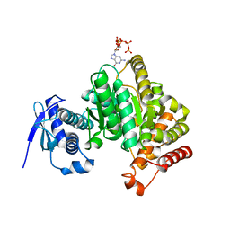
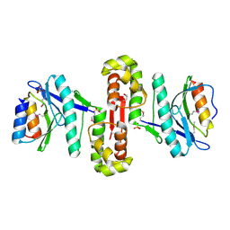
3TND
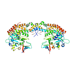
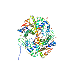 | | Crystal structure of Shigella flexneri VapBC toxin-antitoxin complex | | 分子名称: | Antitoxin VapB, SODIUM ION, SULFATE ION, ... | | 著者 | Dienemann, C, Boggild, A, Winther, K.S, Gerdes, K, Brodersen, D.E. | | 登録日 | 2011-09-01 | | 公開日 | 2011-11-02 | | 最終更新日 | 2024-02-28 | | 実験手法 | X-RAY DIFFRACTION (2.7 Å) | | 主引用文献 | Crystal Structure of the VapBC Toxin-Antitoxin Complex from Shigella flexneri Reveals a Hetero-Octameric DNA-Binding Assembly.
J.Mol.Biol., 414, 2011
|
|
6THH
 | |
7QOC
 | |
6EXP
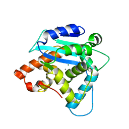
 | | Crystal structure of the SIRV3 AcrID1 (gp02) anti-CRISPR protein | | 分子名称: | SIRV3 AcrID1 (gp02) anti-CRISPR protein | | 著者 | He, F, Bhoobalan-Chitty, Y, Van, L.B, Kjeldsen, A.L, Dedola, M, Makarova, K.S, Koonin, E.V, Brodersen, D.E, Peng, X. | | 登録日 | 2017-11-08 | | 公開日 | 2018-01-31 | | 最終更新日 | 2024-05-08 | | 実験手法 | X-RAY DIFFRACTION (1.93 Å) | | 主引用文献 | Anti-CRISPR proteins encoded by archaeal lytic viruses inhibit subtype I-D immunity.
Nat Microbiol, 3, 2018
|
|
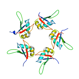
6GFM
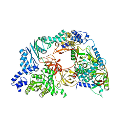
 | | Crystal structure of the Escherichia coli nucleosidase PpnN (pppGpp-form) | | 分子名称: | Pyrimidine/purine nucleotide 5'-monophosphate nucleosidase, guanosine 5'-(tetrahydrogen triphosphate) 3'-(trihydrogen diphosphate) | | 著者 | Zhang, Y, Baerentsen, R.L, Gerdes, K, Brodersen, D.E. | | 登録日 | 2018-05-01 | | 公開日 | 2019-04-24 | | 最終更新日 | 2024-01-17 | | 実験手法 | X-RAY DIFFRACTION (2.77 Å) | | 主引用文献 | (p)ppGpp Regulates a Bacterial Nucleosidase by an Allosteric Two-Domain Switch.
Mol.Cell, 74, 2019
|
|
6GFL
 | |
6GW6
 | |
6HPC
 | |
6HPB
 | |
4V4R
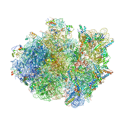 | | Crystal structure of the whole ribosomal complex. | | 分子名称: | 16S ribosomal RNA, 23S ribosomal RNA, 30S ribosomal protein S10, ... | | 著者 | Petry, S, Brodersen, D.E, Murphy IV, F.V, Dunham, C.M, Selmer, M, Tarry, M.J, Kelley, A.C, Ramakrishnan, V. | | 登録日 | 2005-09-30 | | 公開日 | 2014-07-09 | | 最終更新日 | 2024-12-25 | | 実験手法 | X-RAY DIFFRACTION (5.9 Å) | | 主引用文献 | Crystal Structures of the Ribosome in Complex with Release Factors RF1 and RF2 Bound to a Cognate Stop Codon.
Cell(Cambridge,Mass.), 123, 2005
|
|
4V4S
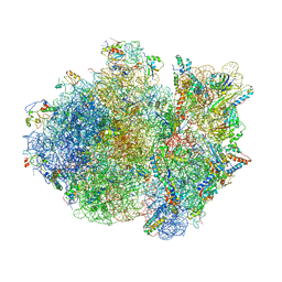 | | Crystal structure of the whole ribosomal complex. | | 分子名称: | 16S ribosomal RNA, 23S ribosomal RNA, 30S ribosomal protein S10, ... | | 著者 | Petry, S, Brodersen, D.E, Murphy IV, F.V, Dunham, C.M, Selmer, M, Tarry, M.J, Kelley, A.C, Ramakrishnan, V. | | 登録日 | 2005-10-12 | | 公開日 | 2014-07-09 | | 最終更新日 | 2024-12-25 | | 実験手法 | X-RAY DIFFRACTION (6.76 Å) | | 主引用文献 | Crystal Structures of the Ribosome in Complex with Release Factors RF1 and RF2 Bound to a Cognate Stop Codon.
Cell(Cambridge,Mass.), 123, 2005
|
|
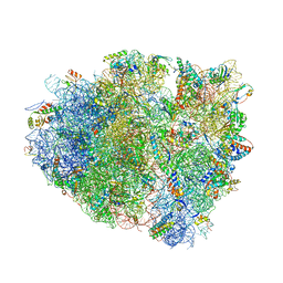
4V7J
 | | Structure of RelE nuclease bound to the 70S ribosome (precleavage state) | | 分子名称: | 30S ribosomal protein S10, 30S ribosomal protein S11, 30S ribosomal protein S12, ... | | 著者 | Neubauer, C, Gao, Y.-G, Andersen, K.R, Dunham, C.M, Kelley, A.C, Hentschel, J, Gerdes, K, Ramakrishnan, V, Brodersen, D.E. | | 登録日 | 2009-11-02 | | 公開日 | 2014-07-09 | | 最終更新日 | 2024-10-16 | | 実験手法 | X-RAY DIFFRACTION (3.3 Å) | | 主引用文献 | The structural basis for mRNA recognition and cleavage by the ribosome-dependent endonuclease RelE.
Cell(Cambridge,Mass.), 139, 2009
|
|
4V7K
 | | Structure of RelE nuclease bound to the 70S ribosome (postcleavage state) | | 分子名称: | 30S ribosomal protein S10, 30S ribosomal protein S11, 30S ribosomal protein S12, ... | | 著者 | Neubauer, C, Gao, Y.-G, Andersen, K.R, Dunham, C.M, Kelley, A.C, Hentschel, J, Gerdes, K, Ramakrishnan, V, Brodersen, D.E. | | 登録日 | 2009-11-02 | | 公開日 | 2014-07-09 | | 最終更新日 | 2024-11-06 | | 実験手法 | X-RAY DIFFRACTION (3.6 Å) | | 主引用文献 | The structural basis for mRNA recognition and cleavage by the ribosome-dependent endonuclease RelE.
Cell(Cambridge,Mass.), 139, 2009
|
|
5IQQ
 | |
3G10
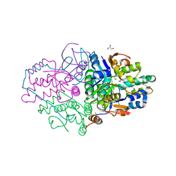
 | | Structure of S. pombe Pop2p - Mg2+ and Mn2+ bound form | | 分子名称: | CCR4-Not complex subunit Caf1, MAGNESIUM ION, MANGANESE (II) ION | | 著者 | Andersen, K.R, Jonstrup, A.T, Van, L.B, Brodersen, D.E. | | 登録日 | 2009-01-29 | | 公開日 | 2009-03-31 | | 最終更新日 | 2023-11-01 | | 実験手法 | X-RAY DIFFRACTION (2.597 Å) | | 主引用文献 | The activity and selectivity of fission yeast Pop2p are affected by a high affinity for Zn2+ and Mn2+ in the active site
Rna, 15, 2009
|
|
4R71
 | | Structure of the Qbeta holoenzyme complex in the P1211 crystal form | | 分子名称: | 30S ribosomal protein S1, Elongation factor Ts, Elongation factor Tu, ... | | 著者 | Gytz, H, Seweryn, P, Kutlubaeva, Z, Chetverin, A.B, Brodersen, D.E, Knudsen, C.R. | | 登録日 | 2014-08-26 | | 公開日 | 2015-09-23 | | 最終更新日 | 2024-02-28 | | 実験手法 | X-RAY DIFFRACTION (3.21 Å) | | 主引用文献 | Structural basis for RNA-genome recognition during bacteriophage Q beta replication.
Nucleic Acids Res., 43, 2015
|
|
5K8J
 | | Structure of Caulobacter crescentus VapBC1 (apo form) | | 分子名称: | GLYCEROL, Ribonuclease VapC, VapB family protein | | 著者 | Bendtsen, K.L, Xu, K, Luckmann, M, Brodersen, D.E. | | 登録日 | 2016-05-30 | | 公開日 | 2016-12-28 | | 最終更新日 | 2024-11-06 | | 実験手法 | X-RAY DIFFRACTION (1.6 Å) | | 主引用文献 | Toxin inhibition in C. crescentus VapBC1 is mediated by a flexible pseudo-palindromic protein motif and modulated by DNA binding.
Nucleic Acids Res., 45, 2017
|
|
5L6L
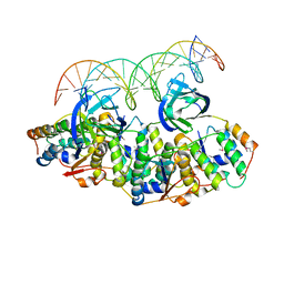 | | Structure of Caulobacter crescentus VapBC1 bound to operator DNA | | 分子名称: | DNA (27-MER), Ribonuclease VapC, VapB family protein | | 著者 | Bendtsen, K.L, Xu, K, Luckmann, M, Brodersen, D.E. | | 登録日 | 2016-05-30 | | 公開日 | 2016-12-28 | | 最終更新日 | 2024-10-16 | | 実験手法 | X-RAY DIFFRACTION (2.7 Å) | | 主引用文献 | Toxin inhibition in C. crescentus VapBC1 is mediated by a flexible pseudo-palindromic protein motif and modulated by DNA binding.
Nucleic Acids Res., 45, 2017
|
|
5L6M
 | | Structure of Caulobacter crescentus VapBC1 (VapB1deltaC:VapC1 form) | | 分子名称: | GLYCEROL, MALONATE ION, Ribonuclease VapC, ... | | 著者 | Bendtsen, K.L, Xu, K, Luckmann, M, Brodersen, D.E. | | 登録日 | 2016-05-30 | | 公開日 | 2016-12-28 | | 最終更新日 | 2024-01-10 | | 実験手法 | X-RAY DIFFRACTION (1.9 Å) | | 主引用文献 | Toxin inhibition in C. crescentus VapBC1 is mediated by a flexible pseudo-palindromic protein motif and modulated by DNA binding.
Nucleic Acids Res., 45, 2017
|
|
7AB5
 | |
7AB4
 | |
7AB3
 | |


