Loading
PDBj
メニューPDBj@FacebookPDBj@TwitterPDBj@YouTubewwPDB FoundationwwPDB
RCSB PDBPDBeBMRBAdv. SearchSearch help
Search by PDB author
4EZR
DownloadVisualize
BU of 4ezr by Molmil
Crystal structure of the substrate binding domain of E.coli DnaK in complex with the C-terminal part of drosocin (residues 12 to 19)
分子名称: Chaperone protein DnaK, Drosocin
著者Zahn, M, Straeter, N.
登録日2012-05-03
公開日2013-04-17
最終更新日2024-02-28
実験手法X-RAY DIFFRACTION (1.9 Å)
主引用文献Structural Studies on the Forward and Reverse Binding Modes of Peptides to the Chaperone DnaK.
J.Mol.Biol., 425, 2013
4EZW
DownloadVisualize
BU of 4ezw by Molmil
Crystal structure of the substrate binding domain of E.coli DnaK in complex with the designer peptide NRLLLTG
分子名称: Chaperone protein DnaK, SULFATE ION, synthetic peptide NRLLLTG
著者Zahn, M, Straeter, N.
登録日2012-05-03
公開日2013-04-17
最終更新日2024-02-28
実験手法X-RAY DIFFRACTION (1.8 Å)
主引用文献Structural Studies on the Forward and Reverse Binding Modes of Peptides to the Chaperone DnaK.
J.Mol.Biol., 425, 2013
4EZO
DownloadVisualize
BU of 4ezo by Molmil
Crystal structure of the substrate binding domain of E.coli DnaK in complex with PR-39 (residues 1 to 15)
分子名称: Antibacterial protein PR-39, Chaperone protein DnaK, SULFATE ION
著者Zahn, M, Straeter, N.
登録日2012-05-03
公開日2013-04-17
最終更新日2024-02-28
実験手法X-RAY DIFFRACTION (1.9 Å)
主引用文献Structural Studies on the Forward and Reverse Binding Modes of Peptides to the Chaperone DnaK.
J.Mol.Biol., 425, 2013
4EZZ
DownloadVisualize
BU of 4ezz by Molmil
Crystal structure of the substrate binding domain of E.coli DnaK in complex with the designer peptide ELPLVKI
分子名称: Chaperone protein DnaK, synthetic peptide ELPLVKI
著者Zahn, M, Straeter, N.
登録日2012-05-03
公開日2013-04-17
最終更新日2024-02-28
実験手法X-RAY DIFFRACTION (2.05 Å)
主引用文献Structural Studies on the Forward and Reverse Binding Modes of Peptides to the Chaperone DnaK.
J.Mol.Biol., 425, 2013
4F00
DownloadVisualize
BU of 4f00 by Molmil
Crystal structure of the substrate binding domain of E.coli DnaK in complex with an apidaecin fragment from the bumblebee (residues 3 to 11)
分子名称: Apidaecin, Chaperone protein DnaK
著者Zahn, M, Straeter, N.
登録日2012-05-03
公開日2013-04-17
最終更新日2024-02-28
実験手法X-RAY DIFFRACTION (1.95 Å)
主引用文献Structural Studies on the Forward and Reverse Binding Modes of Peptides to the Chaperone DnaK.
J.Mol.Biol., 425, 2013
4EZQ
DownloadVisualize
BU of 4ezq by Molmil
Crystal structure of the substrate binding domain of E.coli DnaK in complex with the C-terminal part of pyrrhocoricin (residues 12 to 20)
分子名称: Chaperone protein DnaK, Pyrrhocoricin
著者Zahn, M, Straeter, N.
登録日2012-05-03
公開日2013-04-17
最終更新日2024-02-28
実験手法X-RAY DIFFRACTION (2 Å)
主引用文献Structural Studies on the Forward and Reverse Binding Modes of Peptides to the Chaperone DnaK.
J.Mol.Biol., 425, 2013
4EZU
DownloadVisualize
BU of 4ezu by Molmil
Crystal structure of the substrate binding domain of E.coli DnaK in complex with PR-bombesin in space group I222
分子名称: Chaperone protein DnaK, Proline rich bombesin-related protein
著者Zahn, M, Straeter, N.
登録日2012-05-03
公開日2013-04-24
最終更新日2024-02-28
実験手法X-RAY DIFFRACTION (1.9 Å)
主引用文献Structural studies of DnaK in complex with proline rich antimicrobial peptides reveal two different peptide binding modes
To be Published
4EZS
DownloadVisualize
BU of 4ezs by Molmil
Crystal structure of the substrate binding domain of E.coli DnaK in complex with metchnikowin (residues 20 to 26)
分子名称: Chaperone protein DnaK, Metchnikowin, SULFATE ION
著者Zahn, M, Straeter, N.
登録日2012-05-03
公開日2013-04-24
最終更新日2024-02-28
実験手法X-RAY DIFFRACTION (1.9 Å)
主引用文献Structural studies of DnaK in complex with proline rich antimicrobial peptides reveal two different peptide binding modes
To be Published
4EZY
DownloadVisualize
BU of 4ezy by Molmil
Crystal structure of the substrate binding domain of E.coli DnaK in complex with the designer peptide NRLILTG
分子名称: Chaperone protein DnaK, SULFATE ION, synthetic peptide NRLILTG
著者Zahn, M, Straeter, N.
登録日2012-05-03
公開日2013-04-17
最終更新日2024-02-28
実験手法X-RAY DIFFRACTION (1.85 Å)
主引用文献Structural Studies on the Forward and Reverse Binding Modes of Peptides to the Chaperone DnaK.
J.Mol.Biol., 425, 2013
4EZN
DownloadVisualize
BU of 4ezn by Molmil
Crystal structure of the substrate binding domain of E.coli DnaK in complex with pyrrhocoricin
分子名称: Chaperone protein DnaK, Pyrrhocoricin
著者Zahn, M, Straeter, N.
登録日2012-05-03
公開日2013-04-17
最終更新日2024-02-28
実験手法X-RAY DIFFRACTION (1.8 Å)
主引用文献Structural Studies on the Forward and Reverse Binding Modes of Peptides to the Chaperone DnaK.
J.Mol.Biol., 425, 2013
4EZX
DownloadVisualize
BU of 4ezx by Molmil
Crystal structure of the substrate binding domain of E.coli DnaK in complex with the designer peptide NRLMLTG
分子名称: Chaperone protein DnaK, SULFATE ION, synthetic peptide NRLMLTG
著者Zahn, M, Straeter, N.
登録日2012-05-03
公開日2013-04-17
最終更新日2024-02-28
実験手法X-RAY DIFFRACTION (1.7 Å)
主引用文献Structural Studies on the Forward and Reverse Binding Modes of Peptides to the Chaperone DnaK.
J.Mol.Biol., 425, 2013
4EZV
DownloadVisualize
BU of 4ezv by Molmil
Crystal structure of the substrate binding domain of E.coli DnaK in complex with PR-bombesin in space group P21212
分子名称: Chaperone protein DnaK, Proline rich bombesin-related protein
著者Zahn, M, Straeter, N.
登録日2012-05-03
公開日2013-04-24
最終更新日2024-02-28
実験手法X-RAY DIFFRACTION (2.1 Å)
主引用文献Structural studies of DnaK in complex with proline rich antimicrobial peptides reveal two different peptide binding modes
To be Published
8C65
DownloadVisualize
BU of 8c65 by Molmil
Crystal structure of cutinase AdCut from Acidovorax delafieldii (PBS depolymerase)
分子名称: PBS(A) depolymerase
著者Zahn, M, Clark, M.
登録日2023-01-11
公開日2024-01-24
実験手法X-RAY DIFFRACTION (1.491 Å)
主引用文献Predictions of the substrate specificities of the plastic-degrading enzymes IsPETase and AdCut
To Be Published
3QNJ
DownloadVisualize
BU of 3qnj by Molmil
Crystal structure of the substrate binding domain of E.coli DnaK in complex with the antimicrobial peptide oncocin
分子名称: Chaperone protein DnaK, SULFATE ION, antimicrobial peptide oncocin
著者Zahn, M, Straeter, N.
登録日2011-02-08
公開日2011-03-23
最終更新日2024-02-21
実験手法X-RAY DIFFRACTION (2.28 Å)
主引用文献Rational Design of Oncocin Derivatives with Superior Protease Stabilities and Antibacterial Activities Based on the High-Resolution Structure of the Oncocin-DnaK Complex.
Chembiochem, 12, 2011
4R3U
DownloadVisualize
BU of 4r3u by Molmil
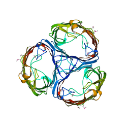
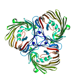
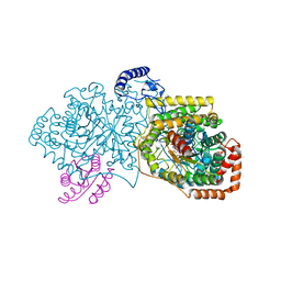
Crystal structure of 2-Hydroxyisobutyryl-CoA Mutase
分子名称: 2-hydroxyisobutyryl-CoA mutase large subunit, 2-hydroxyisobutyryl-CoA mutase small subunit, 3-HYDROXYBUTANOYL-COENZYME A, ...
著者Zahn, M, Kurteva-Yaneva, N, Rohwerder, T, Straeter, N.
登録日2014-08-18
公開日2015-03-11
最終更新日2024-02-28
実験手法X-RAY DIFFRACTION (2.5 Å)
主引用文献Structural basis of the stereospecificity of bacterial B12-dependent 2-hydroxyisobutyryl-CoA mutase.
J.Biol.Chem., 290, 2015
5MDP
DownloadVisualize
BU of 5mdp by Molmil
Crystal structure of in vitro folded Chitoporin VhChip from Vibrio harveyi (crystal form II)
分子名称: Chitoporin
著者Zahn, M, van den Berg, B.
登録日2016-11-13
公開日2017-12-20
最終更新日2024-01-17
実験手法X-RAY DIFFRACTION (3.08 Å)
主引用文献Structural basis for chitin acquisition by marine Vibrio species.
Nat Commun, 9, 2018
8AYV
DownloadVisualize
BU of 8ayv by Molmil
Crystal structure of the Malonyl-ACP Decarboxylase MadB from Pseudomonas putida
分子名称: YiiD_C domain-containing protein
著者Zahn, M, Kuatsjah, E, Beckham, G.T, McGeehan, J.E.
登録日2022-09-03
公開日2023-03-01
最終更新日2024-05-01
実験手法X-RAY DIFFRACTION (1.044 Å)
主引用文献Initiation of fatty acid biosynthesis in Pseudomonas putida KT2440.
Metab Eng, 76, 2023
6EUS
DownloadVisualize
BU of 6eus by Molmil
Crystal structure of the outer membrane channel DcaP of Acinetobacter baumannii
分子名称: DcaP-like protein
著者Zahn, M, van den Berg, B.
登録日2017-10-31
公開日2018-11-14
最終更新日2019-06-19
実験手法X-RAY DIFFRACTION (2.2 Å)
主引用文献A Multidisciplinary Approach toward Identification of Antibiotic Scaffolds for Acinetobacter baumannii.
Structure, 27, 2019
8AIT
DownloadVisualize
BU of 8ait by Molmil
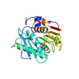
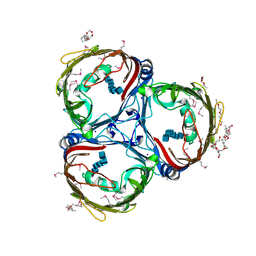
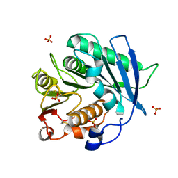
Crystal structure of cutinase PbauzCut from Pseudomonas bauzanensis
分子名称: Cutinase, SULFATE ION
著者Zahn, M, Allen, M.D, Pickford, A.R, McGeehan, J.E.
登録日2022-07-27
公開日2023-03-08
最終更新日2024-02-07
実験手法X-RAY DIFFRACTION (1.24 Å)
主引用文献Concentration-Dependent Inhibition of Mesophilic PETases on Poly(ethylene terephthalate) Can Be Eliminated by Enzyme Engineering.
ChemSusChem, 16, 2023
8AIR
DownloadVisualize
BU of 8air by Molmil
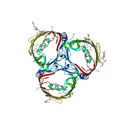
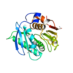
Crystal structure of cutinase RgCutII from Rhizobacter gummiphilus
分子名称: ACETATE ION, RgCutII
著者Zahn, M, Allen, M.D, Pickford, A.R, McGeehan, J.E.
登録日2022-07-27
公開日2023-03-08
最終更新日2024-02-07
実験手法X-RAY DIFFRACTION (1.08 Å)
主引用文献Concentration-Dependent Inhibition of Mesophilic PETases on Poly(ethylene terephthalate) Can Be Eliminated by Enzyme Engineering.
ChemSusChem, 16, 2023
8AIS
DownloadVisualize
BU of 8ais by Molmil
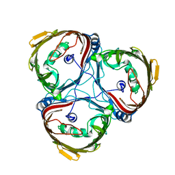
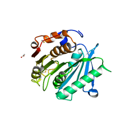
Crystal structure of cutinase PsCut from Pseudomonas saudimassiliensis
分子名称: ACETATE ION, Lipase 1
著者Zahn, M, Allen, M.D, Pickford, A.R, McGeehan, J.E.
登録日2022-07-27
公開日2023-03-08
最終更新日2024-02-07
実験手法X-RAY DIFFRACTION (1.56 Å)
主引用文献Concentration-Dependent Inhibition of Mesophilic PETases on Poly(ethylene terephthalate) Can Be Eliminated by Enzyme Engineering.
ChemSusChem, 16, 2023
5MDS
DownloadVisualize
BU of 5mds by Molmil
Crystal structure of outer membrane expressed Chitoporin VhChip from Vibrio harveyi in complex with chitotetraose
分子名称: 2-acetamido-2-deoxy-beta-D-glucopyranose-(1-4)-2-acetamido-2-deoxy-beta-D-glucopyranose-(1-4)-2-acetamido-2-deoxy-beta-D-glucopyranose-(1-4)-2-acetamido-2-deoxy-beta-D-glucopyranose, Chitoporin
著者Zahn, M, van den Berg, B.
登録日2016-11-13
公開日2017-12-20
最終更新日2024-01-17
実験手法X-RAY DIFFRACTION (2.6 Å)
主引用文献Structural basis for chitin acquisition by marine Vibrio species.
Nat Commun, 9, 2018
5MDR
DownloadVisualize
BU of 5mdr by Molmil
Crystal structure of in vitro folded Chitoporin VhChip from Vibrio harveyi in complex with chitohexaose
分子名称: (HYDROXYETHYLOXY)TRI(ETHYLOXY)OCTANE, 2-acetamido-2-deoxy-beta-D-glucopyranose-(1-4)-2-acetamido-2-deoxy-beta-D-glucopyranose-(1-4)-2-acetamido-2-deoxy-beta-D-glucopyranose-(1-4)-2-acetamido-2-deoxy-beta-D-glucopyranose-(1-4)-2-acetamido-2-deoxy-beta-D-glucopyranose-(1-4)-2-acetamido-2-deoxy-beta-D-glucopyranose, Chitoporin, ...
著者Zahn, M, van den Berg, B.
登録日2016-11-13
公開日2017-12-20
最終更新日2024-01-17
実験手法X-RAY DIFFRACTION (1.9 Å)
主引用文献Structural basis for chitin acquisition by marine Vibrio species.
Nat Commun, 9, 2018
5MDO
DownloadVisualize
BU of 5mdo by Molmil
Crystal structure of in vitro folded Chitoporin VhChip from Vibrio harveyi (crystal form I)
分子名称: (HYDROXYETHYLOXY)TRI(ETHYLOXY)OCTANE, Chitoporin, SODIUM ION
著者Zahn, M, van den Berg, B.
登録日2016-11-13
公開日2017-12-20
最終更新日2018-01-24
実験手法X-RAY DIFFRACTION (1.95 Å)
主引用文献Structural basis for chitin acquisition by marine Vibrio species.
Nat Commun, 9, 2018
5MDQ
DownloadVisualize
BU of 5mdq by Molmil
Crystal structure of outer membrane expressed Chitoporin VhChip from Vibrio harveyi
分子名称: Chitoporin, SODIUM ION
著者Zahn, M, van den Berg, B.
登録日2016-11-13
公開日2017-12-20
最終更新日2024-01-17
実験手法X-RAY DIFFRACTION (2.5 Å)
主引用文献Structural basis for chitin acquisition by marine Vibrio species.
Nat Commun, 9, 2018

220472

件を2024-05-29に公開中

PDB statisticsPDBj update infoContact PDBjnumon