8WFW
 
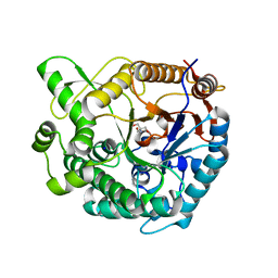 | |
8WFT
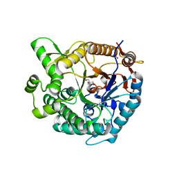 | |
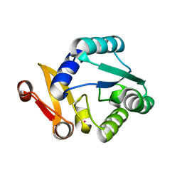
8WFV
 | |
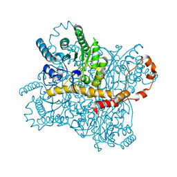
8WGL
 | |
8WDH
 | |
8WGP
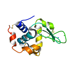
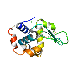
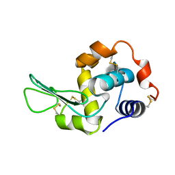
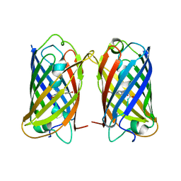 | | Crystal structure of DsRed-Monomer | | 分子名称: | Red fluorescent protein | | 著者 | Nam, K.H. | | 登録日 | 2023-09-22 | | 公開日 | 2024-02-21 | | 実験手法 | X-RAY DIFFRACTION (2.9 Å) | | 主引用文献 | Structural Flexibility of the Monomeric Red Fluorescent Protein DsRed.
Crystals, 14, 2024
|
|
3L1I
 | |
3L1J
 | |
3L1H
 | |
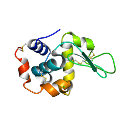
8GMV
 | | Crystal structure of lysozyme | | 分子名称: | CHLORIDE ION, Lysozyme C, SODIUM ION | | 著者 | Nam, K.H. | | 登録日 | 2022-08-22 | | 公開日 | 2022-09-21 | | 最終更新日 | 2023-11-29 | | 実験手法 | X-RAY DIFFRACTION (2.2 Å) | | 主引用文献 | Crystal structure of lysozyme
To Be Published
|
|
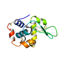
8GMW
 | | Crystal structure of lysozyme | | 分子名称: | CHLORIDE ION, Lysozyme C, SODIUM ION | | 著者 | Nam, K.H. | | 登録日 | 2022-08-22 | | 公開日 | 2022-09-21 | | 最終更新日 | 2023-11-29 | | 実験手法 | X-RAY DIFFRACTION (1.35 Å) | | 主引用文献 | Crystal structure of lysozyme
To Be Published
|
|
8HVF
 | |
8HVE
 | |
7XF7
 | |
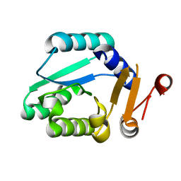
7XF6
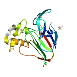
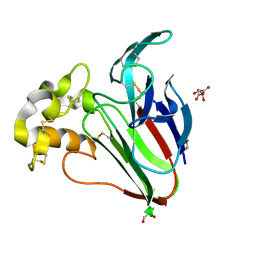
 | | Crystal Structure of Human Lysozyme | | 分子名称: | ACETATE ION, Lysozyme C | | 著者 | Nam, K.H. | | 登録日 | 2022-04-01 | | 公開日 | 2022-04-13 | | 最終更新日 | 2023-11-29 | | 実験手法 | X-RAY DIFFRACTION (1.3 Å) | | 主引用文献 | Crystal Structure of Human Lysozyme Complexed with N-Acetyl-alpha-d-glucosamine.
Appl Sci (Basel), 12, 2022
|
|
7XF8
 | |
7WBE
 | |
7WBF
 | | Crystal structure of lysozyme | | 分子名称: | CHLORIDE ION, Lysozyme C | | 著者 | Nam, K.H. | | 登録日 | 2021-12-16 | | 公開日 | 2022-01-19 | | 最終更新日 | 2023-11-29 | | 実験手法 | X-RAY DIFFRACTION (1.6 Å) | | 主引用文献 | Processing of Multicrystal Diffraction Patterns in Macromolecular Crystallography Using Serial Crystallography Programs.
Crystals, 12, 2022
|
|
7WBD
 | |
6K1W
 | |
6KCC
 | |
6K1X
 | |
6KCA
 | |
6KCD
 | |
6KD1
 | |


