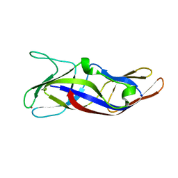
3WGD
 
 | | Crystal structure of ERp46 Trx1 | | 分子名称: | GLYCEROL, PHOSPHATE ION, POTASSIUM ION, ... | | 著者 | Inaba, K, Suzuki, M, Kojima, R. | | 登録日 | 2013-08-04 | | 公開日 | 2014-06-25 | | 実験手法 | X-RAY DIFFRACTION (2.5 Å) | | 主引用文献 | Radically different thioredoxin domain arrangement of ERp46, an efficient disulfide bond introducer of the mammalian PDI family
Structure, 22, 2014
|
|
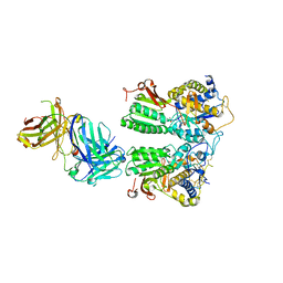
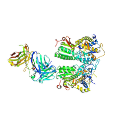
3WGX
 | | Crystal structure of ERp46 Trx2 in a complex with Prx4 C-term | | 分子名称: | GLYCEROL, Peroxiredoxin-4, Thioredoxin domain-containing protein 5 | | 著者 | Inaba, K, Suzuki, M, Kojima, R. | | 登録日 | 2013-08-13 | | 公開日 | 2014-06-25 | | 実験手法 | X-RAY DIFFRACTION (0.92 Å) | | 主引用文献 | Radically different thioredoxin domain arrangement of ERp46, an efficient disulfide bond introducer of the mammalian PDI family
Structure, 22, 2014
|
|
5ZLQ
 | | Crystal Structure of C1orf123 | | 分子名称: | UPF0587 protein C1orf123, ZINC ION | | 著者 | Furukawa, Y, Lim, C.T, Tosha, T. | | 登録日 | 2018-03-29 | | 公開日 | 2018-10-10 | | 最終更新日 | 2024-03-27 | | 実験手法 | X-RAY DIFFRACTION (2 Å) | | 主引用文献 | Identification of a novel zinc-binding protein, C1orf123, as an interactor with a heavy metal-associated domain
PLoS ONE, 13, 2018
|
|
5X2Q
 | | Crystal structure of the medaka fish taste receptor T1r2a-T1r3 ligand binding domains in complex with glycine | | 分子名称: | 2-acetamido-2-deoxy-beta-D-glucopyranose, 2-acetamido-2-deoxy-beta-D-glucopyranose-(1-4)-2-acetamido-2-deoxy-beta-D-glucopyranose, CALCIUM ION, ... | | 著者 | Nuemket, N, Yasui, N, Atsumi, N, Yamashita, A. | | 登録日 | 2017-02-02 | | 公開日 | 2017-05-24 | | 最終更新日 | 2023-11-22 | | 実験手法 | X-RAY DIFFRACTION (2.6 Å) | | 主引用文献 | Structural basis for perception of diverse chemical substances by T1r taste receptors
Nat Commun, 8, 2017
|
|
5X2O
 | | Crystal structure of the medaka fish taste receptor T1r2a-T1r3 ligand binding domains in complex with L-arginine | | 分子名称: | 2-acetamido-2-deoxy-beta-D-glucopyranose, ARGININE, CALCIUM ION, ... | | 著者 | Nuemket, N, Yasui, N, Atsumi, N, Yamashita, A. | | 登録日 | 2017-02-02 | | 公開日 | 2017-05-24 | | 最終更新日 | 2023-11-22 | | 実験手法 | X-RAY DIFFRACTION (2.6 Å) | | 主引用文献 | Structural basis for perception of diverse chemical substances by T1r taste receptors
Nat Commun, 8, 2017
|
|
3A0R
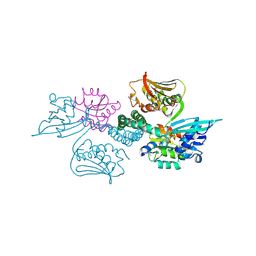 | | Crystal structure of histidine kinase ThkA (TM1359) in complex with response regulator protein TrrA (TM1360) | | 分子名称: | MERCURY (II) ION, Response regulator, Sensor protein | | 著者 | Yamada, S, Sugimoto, H, Kobayashi, M, Ohno, A, Nakamura, H, Shiro, Y. | | 登録日 | 2009-03-24 | | 公開日 | 2009-10-20 | | 最終更新日 | 2021-11-10 | | 実験手法 | X-RAY DIFFRACTION (3.8 Å) | | 主引用文献 | Structure of PAS-linked histidine kinase and the response regulator complex
Structure, 17, 2009
|
|


