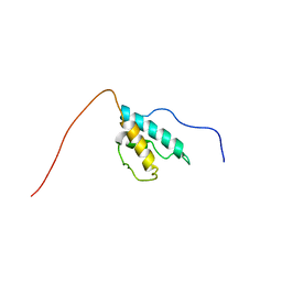
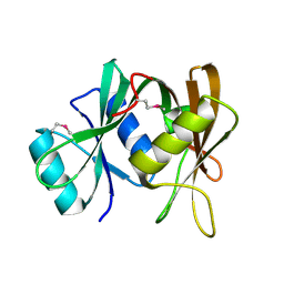
2CQI
 
 | | Solution structure of the RNA binding domain of Nucleolysin TIAR | | 分子名称: | Nucleolysin TIAR | | 著者 | Suzuki, S, Muto, Y, Inoue, M, Kigawa, T, Terada, T, Shirouzu, M, Yokoyama, S, RIKEN Structural Genomics/Proteomics Initiative (RSGI) | | 登録日 | 2005-05-20 | | 公開日 | 2005-11-20 | | 最終更新日 | 2024-05-01 | | 実験手法 | SOLUTION NMR | | 主引用文献 | Solution structure of the RNA binding domain of Nucleolysin TIAR
To be Published
|
|
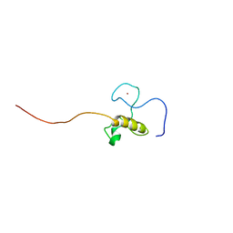
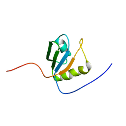
2CPJ
 | | Solution structure of the N-terminal RNA recognition motif of NonO | | 分子名称: | Non-POU domain-containing octamer-binding protein | | 著者 | Nagata, T, Muto, Y, Inoue, M, Kigawa, T, Terada, T, Shirouzu, M, Yokoyama, S, RIKEN Structural Genomics/Proteomics Initiative (RSGI) | | 登録日 | 2005-05-19 | | 公開日 | 2005-11-19 | | 最終更新日 | 2024-05-29 | | 実験手法 | SOLUTION NMR | | 主引用文献 | Solution structure of the N-terminal RNA recognition motif of NonO
To be Published
|
|
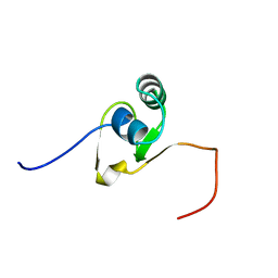
2CQD
 | | Solution Structure of the RNA recognition motif in RNA-binding region containing protein 1 | | 分子名称: | RNA-binding region containing protein 1 | | 著者 | Someya, T, Muto, Y, Inoue, M, Kigawa, T, Terada, T, Shirouzu, M, Yokoyama, S, RIKEN Structural Genomics/Proteomics Initiative (RSGI) | | 登録日 | 2005-05-19 | | 公開日 | 2005-11-19 | | 最終更新日 | 2024-05-29 | | 実験手法 | SOLUTION NMR | | 主引用文献 | Solution Structure of the RNA recognition motif in RNA-binding region containing protein 1
To be Published
|
|
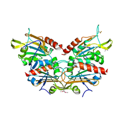
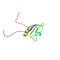
2CZ4
 | | Crystal structure of a putative PII-like signaling protein (TTHA0516) from Thermus thermophilus HB8 | | 分子名称: | ACETATE ION, CHLORIDE ION, hypothetical protein TTHA0516 | | 著者 | Arai, R, Fusatomi, E, Kukimoto-Niino, M, Kawaguchi, S, Terada, T, Shirouzu, M, Yokoyama, S, RIKEN Structural Genomics/Proteomics Initiative (RSGI) | | 登録日 | 2005-07-10 | | 公開日 | 2006-01-10 | | 最終更新日 | 2024-11-13 | | 実験手法 | X-RAY DIFFRACTION (1.93 Å) | | 主引用文献 | Crystal structure of a putative PII-like signaling protein (TTHA0516) from Thermus thermophilus HB8
To be Published
|
|
2CYE
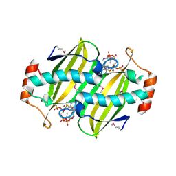 | | Crystal structure of Thioesterase complexed with coenzyme A and Zn from Thermus thermophilus HB8 | | 分子名称: | COENZYME A, Putative thioesterase, ZINC ION | | 著者 | Hosaka, T, Kato-Murayama, M, Murayama, K, Kishishita, S, Handa, N, Shirouzu, M, Yokoyama, S, RIKEN Structural Genomics/Proteomics Initiative (RSGI) | | 登録日 | 2005-07-06 | | 公開日 | 2006-01-06 | | 最終更新日 | 2024-10-30 | | 実験手法 | X-RAY DIFFRACTION (1.9 Å) | | 主引用文献 | Structure of the putative thioesterase protein TTHA1846 from Thermus thermophilus HB8 complexed with coenzyme A and a zinc ion.
Acta Crystallogr.,Sect.D, 65, 2009
|
|
2CPR
 | | Solution structure of the HRDC domain of human Exosome component 10 | | 分子名称: | Exosome component 10 | | 著者 | Nagata, T, Muto, Y, Inoue, M, Kigawa, T, Terada, T, Shirouzu, M, Yokoyama, S, RIKEN Structural Genomics/Proteomics Initiative (RSGI) | | 登録日 | 2005-05-19 | | 公開日 | 2005-11-19 | | 最終更新日 | 2024-05-29 | | 実験手法 | SOLUTION NMR | | 主引用文献 | Solution structure of the HRDC domain of human Exosome component 10
To be Published
|
|
2CQE
 | | Solution Structure of the Zinc-finger domain in KIAA1064 protein | | 分子名称: | KIAA1064 protein, ZINC ION | | 著者 | Someya, T, Muto, Y, Inoue, M, Kigawa, T, Terada, T, Shirouzu, M, Yokoyama, S, RIKEN Structural Genomics/Proteomics Initiative (RSGI) | | 登録日 | 2005-05-19 | | 公開日 | 2005-11-19 | | 最終更新日 | 2024-05-29 | | 実験手法 | SOLUTION NMR | | 主引用文献 | Solution Structure of the Zinc-finger domain in KIAA1064 protein
To be Published
|
|
2CX3
 | | Crystal structure of a bacterioferritin comigratory protein peroxiredoxin from the Aeropyrum pernix K1 (form-1 crystal) | | 分子名称: | bacterioferritin comigratory protein | | 著者 | Mizohata, E, Murayama, K, Idaka, M, Tatsuguchi, A, Terada, T, Shirouzu, M, Yokoyama, S, RIKEN Structural Genomics/Proteomics Initiative (RSGI) | | 登録日 | 2005-06-27 | | 公開日 | 2005-12-27 | | 最終更新日 | 2024-10-09 | | 実験手法 | X-RAY DIFFRACTION (2.64 Å) | | 主引用文献 | Crystal structure of a bacterioferritin comigratory protein peroxiredoxin from the Aeropyrum pernix K1 (form-1 crystal)
To be Published
|
|
2CXH
 | | Crystal structure of probable ribosomal biogenesis protein from Aeropyrum pernix K1 | | 分子名称: | Probable brix-domain ribosomal biogenesis protein | | 著者 | Kawazoe, M, Takemoto, C, Hanawa-Suetsugu, K, Kaminishi, T, Terada, T, Shirouzu, M, Yokoyama, S, RIKEN Structural Genomics/Proteomics Initiative (RSGI) | | 登録日 | 2005-06-29 | | 公開日 | 2005-12-29 | | 最終更新日 | 2024-11-13 | | 実験手法 | X-RAY DIFFRACTION (1.8 Å) | | 主引用文献 | Crystal structure of probable ribosomal biogenesis protein from Aeropyrum pernix K1
To be Published
|
|
2CQ1
 | | solution structure of RNA binding domain in PTB-like protein L | | 分子名称: | PTB-like protein L | | 著者 | Tsuda, K, Muto, Y, Inoue, M, Kigawa, T, Terada, T, Shirouzu, M, Yokoyama, S, RIKEN Structural Genomics/Proteomics Initiative (RSGI) | | 登録日 | 2005-05-19 | | 公開日 | 2005-11-19 | | 最終更新日 | 2024-05-29 | | 実験手法 | SOLUTION NMR | | 主引用文献 | solution structure of RNA binding domain in PTB-like protein L
To be Published
|
|
2CQJ
 | | Solution structure of the S4 domain of U3 small nucleolar ribonucleoprotein protein IMP3 homolog | | 分子名称: | U3 small nucleolar ribonucleoprotein protein IMP3 homolog | | 著者 | Suzuki, S, Muto, Y, Inoue, M, Kigawa, T, Terada, T, Shirouzu, M, Yokoyama, S, RIKEN Structural Genomics/Proteomics Initiative (RSGI) | | 登録日 | 2005-05-20 | | 公開日 | 2005-11-20 | | 最終更新日 | 2024-05-29 | | 実験手法 | SOLUTION NMR | | 主引用文献 | Solution structure of the S4 domain of U3 small nucleolar ribonucleoprotein protein IMP3 homolog
To be Published
|
|
2CPE
 | | Solution structure of the RNA recognition motif of Ewing Sarcoma(EWS) protein | | 分子名称: | RNA-binding protein EWS | | 著者 | Nagata, T, Muto, Y, Inoue, M, Kigawa, T, Terada, T, Shirouzu, M, Yokoyama, S, RIKEN Structural Genomics/Proteomics Initiative (RSGI) | | 登録日 | 2005-05-19 | | 公開日 | 2005-11-19 | | 最終更新日 | 2024-05-29 | | 実験手法 | SOLUTION NMR | | 主引用文献 | Solution structure of the RNA recognition motif of Ewing Sarcoma(EWS) protein
To be Published
|
|
2CQ0
 | | solution structure of RNA binding domain in eukaryotic translation initiation factor 3 subunit 4 | | 分子名称: | Eukaryotic translation initiation factor 3 subunit 4 | | 著者 | Tsuda, K, Muto, Y, Inoue, M, Kigawa, T, Terada, T, Shirouzu, M, Yokoyama, S, RIKEN Structural Genomics/Proteomics Initiative (RSGI) | | 登録日 | 2005-05-19 | | 公開日 | 2005-11-19 | | 最終更新日 | 2024-05-29 | | 実験手法 | SOLUTION NMR | | 主引用文献 | solution structure of RNA binding domain in eukaryotic translation initiation factor 3 subunit 4
To be Published
|
|
2CQK
 | | Solution structure of the La domain of c-Mpl binding protein | | 分子名称: | C-Mpl binding protein | | 著者 | Suzuki, S, Muto, Y, Inoue, M, Kigawa, T, Terada, T, Shirouzu, M, Yokoyama, S, RIKEN Structural Genomics/Proteomics Initiative (RSGI) | | 登録日 | 2005-05-20 | | 公開日 | 2005-11-20 | | 最終更新日 | 2024-05-29 | | 実験手法 | SOLUTION NMR | | 主引用文献 | Solution structure of the La domain of c-Mpl binding protein
To be Published
|
|
2RS7
 | | Solution structure of the second dsRBD from RNA helicase A | | 分子名称: | ATP-dependent RNA helicase A | | 著者 | Nagata, T, Muto, Y, Tsuda, K, Inoue, M, Kigawa, T, Terada, T, Shirouzu, M, Yokoyama, S, RIKEN Structural Genomics/Proteomics Initiative (RSGI) | | 登録日 | 2011-11-29 | | 公開日 | 2012-03-14 | | 最終更新日 | 2024-05-15 | | 実験手法 | SOLUTION NMR | | 主引用文献 | Solution structures of the double-stranded RNA-binding domains from RNA helicase A
Proteins, 80, 2012
|
|
2RS6
 | | Solution structure of the N-terminal dsRBD from RNA helicase A | | 分子名称: | ATP-dependent RNA helicase A | | 著者 | Nagata, T, Muto, Y, Tsuda, K, Inoue, M, Kigawa, T, Terada, T, Shirouzu, M, Yokoyama, S, RIKEN Structural Genomics/Proteomics Initiative (RSGI) | | 登録日 | 2011-11-29 | | 公開日 | 2012-03-14 | | 最終更新日 | 2024-05-15 | | 実験手法 | SOLUTION NMR | | 主引用文献 | Solution structures of the double-stranded RNA-binding domains from RNA helicase A
Proteins, 80, 2012
|
|
2CPQ
 | | Solution structure of the N-terminal KH domain of human FXR1 | | 分子名称: | Fragile X mental retardation syndrome related protein 1, isoform b' | | 著者 | Nagata, T, Muto, Y, Inoue, M, Kigawa, T, Terada, T, Shirouzu, M, Yokoyama, S, RIKEN Structural Genomics/Proteomics Initiative (RSGI) | | 登録日 | 2005-05-19 | | 公開日 | 2005-11-19 | | 最終更新日 | 2024-05-29 | | 実験手法 | SOLUTION NMR | | 主引用文献 | Solution structure of the N-terminal KH domain of human FXR1
To be Published
|
|
2CQB
 | | Solution Structure of the RNA recognition motif in Peptidyl-prolyl cis-trans isomerase E | | 分子名称: | Peptidyl-prolyl cis-trans isomerase E | | 著者 | Someya, T, Muto, Y, Inoue, M, Kigawa, T, Terada, T, Shirouzu, M, Yokoyama, S, RIKEN Structural Genomics/Proteomics Initiative (RSGI) | | 登録日 | 2005-05-19 | | 公開日 | 2005-11-19 | | 最終更新日 | 2024-05-29 | | 実験手法 | SOLUTION NMR | | 主引用文献 | Solution Structure of the RNA recognition motif in Peptidyl-prolyl cis-trans isomerase E
To be Published
|
|
2CWP
 | |
2CPI
 | | Solution structure of the RNA recognition motif of CNOT4 | | 分子名称: | CCR4-NOT transcription complex subunit 4 | | 著者 | Nagata, T, Muto, Y, Inoue, M, Kigawa, T, Terada, T, Shirouzu, M, Yokoyama, S, RIKEN Structural Genomics/Proteomics Initiative (RSGI) | | 登録日 | 2005-05-19 | | 公開日 | 2005-11-19 | | 最終更新日 | 2024-05-29 | | 実験手法 | SOLUTION NMR | | 主引用文献 | Solution structure of the RNA recognition motif of CNOT4
To be Published
|
|
2CQL
 | | Solution structure of the N-terminal domain of human ribosomal protein L9 | | 分子名称: | 60S ribosomal protein L9 | | 著者 | Suzuki, S, Muto, Y, Inoue, M, Kigawa, T, Terada, T, Shirouzu, M, Yokoyama, S, RIKEN Structural Genomics/Proteomics Initiative (RSGI) | | 登録日 | 2005-05-20 | | 公開日 | 2005-11-20 | | 最終更新日 | 2024-05-29 | | 実験手法 | SOLUTION NMR | | 主引用文献 | Solution structure of the N-terminal domain of human ribosomal protein L9
To be Published
|
|
2CPH
 | | Solution structure of the C-terminal RNA recognition motif of hypothetical RNA-binding protein RBM19 | | 分子名称: | RNA binding motif protein 19 | | 著者 | Nagata, T, Muto, Y, Inoue, M, Kigawa, T, Terada, T, Shirouzu, M, Yokoyama, S, RIKEN Structural Genomics/Proteomics Initiative (RSGI) | | 登録日 | 2005-05-19 | | 公開日 | 2005-11-19 | | 最終更新日 | 2024-05-29 | | 実験手法 | SOLUTION NMR | | 主引用文献 | Solution structure of the C-terminal RNA recognition motif of hypothetical RNA-binding protein RBM19
To be Published
|
|
2CQO
 | | Solution structure of the S1 RNA binding domain of human hypothetical protein FLJ11067 | | 分子名称: | Nucleolar protein of 40 kDa | | 著者 | Suzuki, S, Muto, Y, Inoue, M, Kigawa, T, Terada, T, Shirouzu, M, Yokoyama, S, RIKEN Structural Genomics/Proteomics Initiative (RSGI) | | 登録日 | 2005-05-20 | | 公開日 | 2005-11-20 | | 最終更新日 | 2024-05-29 | | 実験手法 | SOLUTION NMR | | 主引用文献 | Solution structure of the S1 RNA binding domain of human hypothetical protein FLJ11067
To be Published
|
|
2CPM
 | | Solution structure of the R3H domain of human sperm-associated antigen 7 | | 分子名称: | Sperm-associated antigen 7 | | 著者 | Nagata, T, Muto, Y, Inoue, M, Kigawa, T, Terada, T, Shirouzu, M, Yokoyama, S, RIKEN Structural Genomics/Proteomics Initiative (RSGI) | | 登録日 | 2005-05-19 | | 公開日 | 2005-11-19 | | 最終更新日 | 2024-05-29 | | 実験手法 | SOLUTION NMR | | 主引用文献 | Solution structure of the R3H domain of human sperm-associated antigen 7
To be Published
|
|
2CQ4
 | | solution structure of RNA binding domain in RNA binding motif protein 23 | | 分子名称: | RNA binding motif protein 23 | | 著者 | Tsuda, K, Muto, Y, Inoue, M, Kigawa, T, Terada, T, Shirouzu, M, Yokoyama, S, RIKEN Structural Genomics/Proteomics Initiative (RSGI) | | 登録日 | 2005-05-19 | | 公開日 | 2005-11-19 | | 最終更新日 | 2024-05-29 | | 実験手法 | SOLUTION NMR | | 主引用文献 | solution structure of RNA binding domain in RNA binding motif protein 23
To be Published
|
|


