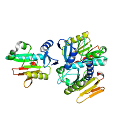
1WCW
 
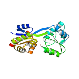 | | Crystal Structure of Uroporphyrinogen III Synthase from an Extremely Thermophilic Bacterium Thermus thermophilus HB8 (Wild type, Native, Form-1 crystal) | | 分子名称: | GLYCEROL, Uroporphyrinogen III synthase | | 著者 | Mizohata, E, Matsuura, T, Sakai, H, Murayama, K, Terada, T, Shirouzu, M, Kuramitsu, S, Yokoyama, S, RIKEN Structural Genomics/Proteomics Initiative (RSGI) | | 登録日 | 2004-05-06 | | 公開日 | 2005-05-06 | | 最終更新日 | 2024-03-13 | | 実験手法 | X-RAY DIFFRACTION (1.3 Å) | | 主引用文献 | Crystal Structure of Uroporphyrinogen III Synthase from Thermus thermophilus HB8
To be Published
|
|
1X4G
 | | Solution structure of RRM domain in Nucleolysin TIAR | | 分子名称: | Nucleolysin TIAR | | 著者 | He, F, Muto, Y, Inoue, M, Kigawa, T, Shirouzu, M, Terada, T, Yokoyama, S, RIKEN Structural Genomics/Proteomics Initiative (RSGI) | | 登録日 | 2005-05-14 | | 公開日 | 2005-11-14 | | 最終更新日 | 2024-05-29 | | 実験手法 | SOLUTION NMR | | 主引用文献 | Solution structure of RRM domain in Nucleolysin TIAR
To be Published
|
|
1WEO
 | | Solution structure of RING-finger in the catalytic subunit (IRX3) of cellulose synthase | | 分子名称: | ZINC ION, cellulose synthase, catalytic subunit (IRX3) | | 著者 | He, F, Muto, Y, Inoue, M, Kigawa, T, Shirouzu, M, Terada, T, Yokoyama, S, RIKEN Structural Genomics/Proteomics Initiative (RSGI) | | 登録日 | 2004-05-25 | | 公開日 | 2004-11-25 | | 最終更新日 | 2024-05-29 | | 実験手法 | SOLUTION NMR | | 主引用文献 | Solution structure of RING-finger in the catalytic subunit (IRX3) of cellulose synthase
To be Published
|
|
1WF0
 | | Solution structure of RRM domain in TAR DNA-binding protein-43 | | 分子名称: | TAR DNA-binding protein-43 | | 著者 | He, F, Muto, Y, Inoue, M, Kigawa, T, Shirouzu, M, Terada, T, Yokoyama, S, RIKEN Structural Genomics/Proteomics Initiative (RSGI) | | 登録日 | 2004-05-25 | | 公開日 | 2004-11-25 | | 最終更新日 | 2024-05-29 | | 実験手法 | SOLUTION NMR | | 主引用文献 | Solution structure of RRM domain in TAR DNA-binding protein-43
To be Published
|
|
1WIB
 | | Solution structure of the N-terminal domain from mouse hypothetical protein BAB22488 | | 分子名称: | 60S ribosomal protein L12 | | 著者 | Suzuki, S, Muto, Y, Inoue, M, Kigawa, T, Terada, T, Shirouzu, M, Yokoyama, S, RIKEN Structural Genomics/Proteomics Initiative (RSGI) | | 登録日 | 2004-05-28 | | 公開日 | 2004-11-28 | | 最終更新日 | 2024-05-29 | | 実験手法 | SOLUTION NMR | | 主引用文献 | Solution structure of the N-terminal domain from mouse hypothetical protein BAB22488
To be Published
|
|
1X4E
 | | Solution structure of RRM domain in RNA binding motif, single-stranded interacting protein 2 | | 分子名称: | RNA binding motif, single-stranded interacting protein 2 | | 著者 | He, F, Muto, Y, Inoue, M, Kigawa, T, Shirouzu, M, Terada, T, Yokoyama, S, RIKEN Structural Genomics/Proteomics Initiative (RSGI) | | 登録日 | 2005-05-14 | | 公開日 | 2005-11-14 | | 最終更新日 | 2024-05-29 | | 実験手法 | SOLUTION NMR | | 主引用文献 | Solution structure of RRM domain in RNA binding motif, single-stranded interacting protein 2
To be Published
|
|
1WE8
 | | Solution structure of KH domain in protein BAB28342 | | 分子名称: | Tudor and KH domain containing protein | | 著者 | He, F, Muto, Y, Inoue, M, Kigawa, T, Shirouzu, M, Terada, T, Hayashi, F, Yokoyama, S, RIKEN Structural Genomics/Proteomics Initiative (RSGI) | | 登録日 | 2004-05-24 | | 公開日 | 2004-11-24 | | 最終更新日 | 2024-05-29 | | 実験手法 | SOLUTION NMR | | 主引用文献 | Solution structure of KH domain in protein BAB28342
To be Published
|
|
1WEX
 | | Solution structure of RRM domain in protein BAB28521 | | 分子名称: | HYPOTHETICAL PROTEIN (RIKEN CDNA 2810036L13) | | 著者 | He, F, Muto, Y, Inoue, M, Kigawa, T, Shirouzu, M, Terada, T, Yokoyama, S, RIKEN Structural Genomics/Proteomics Initiative (RSGI) | | 登録日 | 2004-05-25 | | 公開日 | 2004-11-25 | | 最終更新日 | 2024-05-29 | | 実験手法 | SOLUTION NMR | | 主引用文献 | Solution structure of RRM domain in protein BAB28521
To be Published
|
|
1X4M
 | | Solution structure of KH domain in Far upstream element binding protein 1 | | 分子名称: | Far upstream element binding protein 1 | | 著者 | He, F, Muto, Y, Inoue, M, Kigawa, T, Shirouzu, M, Terada, T, Yokoyama, S, RIKEN Structural Genomics/Proteomics Initiative (RSGI) | | 登録日 | 2005-05-14 | | 公開日 | 2005-11-14 | | 最終更新日 | 2024-05-29 | | 実験手法 | SOLUTION NMR | | 主引用文献 | Solution structure of KH domain in Far upstream element binding protein 1
To be Published
|
|
1WEE
 | | Solution structure of PHD domain in PHD finger family protein | | 分子名称: | PHD finger family protein, ZINC ION | | 著者 | He, F, Muto, Y, Inoue, M, Kigawa, T, Shirouzu, M, Terada, T, Yokoyama, S, RIKEN Structural Genomics/Proteomics Initiative (RSGI) | | 登録日 | 2004-05-24 | | 公開日 | 2004-11-24 | | 最終更新日 | 2024-05-29 | | 実験手法 | SOLUTION NMR | | 主引用文献 | Solution structure of PHD domain in PHD finger family protein
To be Published
|
|
1X4K
 | | Solution structure of LIM domain in LIM-protein 3 | | 分子名称: | Skeletal muscle LIM-protein 3, ZINC ION | | 著者 | He, F, Muto, Y, Inoue, M, Kigawa, T, Shirouzu, M, Terada, T, Yokoyama, S, RIKEN Structural Genomics/Proteomics Initiative (RSGI) | | 登録日 | 2005-05-14 | | 公開日 | 2005-11-14 | | 最終更新日 | 2024-05-29 | | 実験手法 | SOLUTION NMR | | 主引用文献 | Solution structure of LIM domain in LIM-protein 3
To be Published
|
|
1WG5
 | | Solution structure of the first RRM domain in heterogeneous nuclear ribonucleoprotein H | | 分子名称: | Heterogeneous nuclear ribonucleoprotein H | | 著者 | He, F, Muto, Y, Inoue, M, Kigawa, T, Shirouzu, M, Terada, T, Yokoyama, S, RIKEN Structural Genomics/Proteomics Initiative (RSGI) | | 登録日 | 2004-05-27 | | 公開日 | 2004-11-27 | | 最終更新日 | 2024-05-29 | | 実験手法 | SOLUTION NMR | | 主引用文献 | Solution structure of the first RRM domain in heterogeneous nuclear ribonucleoprotein H
To be Published
|
|
1X4A
 | | Solution structure of RRM domain in splicing factor SF2 | | 分子名称: | splicing factor, arginine/serine-rich 1 (splicing factor 2, alternate splicing factor) variant | | 著者 | He, F, Muto, Y, Inoue, M, Kigawa, T, Shirouzu, M, Terada, T, Yokoyama, S, RIKEN Structural Genomics/Proteomics Initiative (RSGI) | | 登録日 | 2005-05-14 | | 公開日 | 2005-11-14 | | 最終更新日 | 2024-05-29 | | 実験手法 | SOLUTION NMR | | 主引用文献 | Solution structure of RRM domain in splicing factor SF2
To be Published
|
|
1X4Q
 | | Solution structure of PWI domain in U4/U6 small nuclear ribonucleoprotein Prp3(hPrp3) | | 分子名称: | U4/U6 small nuclear ribonucleoprotein Prp3 | | 著者 | He, F, Muto, Y, Inoue, M, Kigawa, T, Shirouzu, M, Terada, T, Yokoyama, S, RIKEN Structural Genomics/Proteomics Initiative (RSGI) | | 登録日 | 2005-05-14 | | 公開日 | 2005-11-14 | | 最終更新日 | 2024-05-29 | | 実験手法 | SOLUTION NMR | | 主引用文献 | Solution structure of PWI domain in U4/U6 small nuclear ribonucleoprotein Prp3(hPrp3)
To be Published
|
|
1X49
 | | Solution structure of the first DSRM domain in Interferon-induced, double-stranded RNA-activated protein kinase | | 分子名称: | Interferon-induced, double-stranded RNA-activated protein kinase | | 著者 | He, F, Muto, Y, Inoue, M, Kigawa, T, Shirouzu, M, Terada, T, Yokoyama, S, RIKEN Structural Genomics/Proteomics Initiative (RSGI) | | 登録日 | 2005-05-14 | | 公開日 | 2005-11-14 | | 最終更新日 | 2024-05-29 | | 実験手法 | SOLUTION NMR | | 主引用文献 | Solution structure of the first DSRM domain in Interferon-induced, double-stranded RNA-activated protein kinase
To be Published
|
|
1X4N
 | | Solution structure of KH domain in FUSE binding protein 1 | | 分子名称: | Far upstream element binding protein 1 | | 著者 | He, F, Muto, Y, Inoue, M, Kigawa, T, Shirouzu, M, Terada, T, Yokoyama, S, RIKEN Structural Genomics/Proteomics Initiative (RSGI) | | 登録日 | 2005-05-14 | | 公開日 | 2005-11-14 | | 最終更新日 | 2024-05-29 | | 実験手法 | SOLUTION NMR | | 主引用文献 | Solution structure of KH domain in FUSE binding protein 1
To be Published
|
|
1X47
 | | Solution structure of DSRM domain in DGCR8 protein | | 分子名称: | DGCR8 protein | | 著者 | He, F, Muto, Y, Inoue, M, Kigawa, T, Shirouzu, M, Terada, T, Yokoyama, S, RIKEN Structural Genomics/Proteomics Initiative (RSGI) | | 登録日 | 2005-05-14 | | 公開日 | 2005-11-14 | | 最終更新日 | 2024-05-29 | | 実験手法 | SOLUTION NMR | | 主引用文献 | Solution structure of DSRM domain in DGCR8 protein
To be Published
|
|
1X4P
 | | Solution structure of SURP domain in SFRS14 protei | | 分子名称: | Putative splicing factor, arginine/serine-rich 14 | | 著者 | He, F, Muto, Y, Inoue, M, Kigawa, T, Shirouzu, M, Terada, T, Yokoyama, S, RIKEN Structural Genomics/Proteomics Initiative (RSGI) | | 登録日 | 2005-05-14 | | 公開日 | 2005-11-14 | | 最終更新日 | 2024-05-29 | | 実験手法 | SOLUTION NMR | | 主引用文献 | Solution structure of SURP domain in SFRS14 protei
To be Published
|
|
1X4I
 | | Solution structure of PHD domain in inhibitor of growth protein 3 (ING3) | | 分子名称: | Inhibitor of growth protein 3, ZINC ION | | 著者 | He, F, Muto, Y, Inoue, M, Kigawa, T, Shirouzu, M, Terada, T, Yokoyama, S, RIKEN Structural Genomics/Proteomics Initiative (RSGI) | | 登録日 | 2005-05-14 | | 公開日 | 2005-11-14 | | 最終更新日 | 2024-05-29 | | 実験手法 | SOLUTION NMR | | 主引用文献 | Solution structure of PHD domain in inhibitor of growth protein 3 (ING3)
To be Published
|
|
1X4R
 | | Solution structure of WWE domain in Parp14 protein | | 分子名称: | Parp14 protein | | 著者 | He, F, Muto, Y, Inoue, M, Kigawa, T, Shirouzu, M, Terada, T, Yokoyama, S, RIKEN Structural Genomics/Proteomics Initiative (RSGI) | | 登録日 | 2005-05-14 | | 公開日 | 2005-11-14 | | 最終更新日 | 2024-05-29 | | 実験手法 | SOLUTION NMR | | 主引用文献 | Solution structure of WWE domain in Parp14 protein
To be Published
|
|
1X5S
 | | Solution structure of RRM domain in A18 hnRNP | | 分子名称: | Cold-inducible RNA-binding protein | | 著者 | Sato, A, Muto, Y, Inoue, M, Kigawa, T, Shirouzu, M, Terada, T, Yokoyama, S, RIKEN Structural Genomics/Proteomics Initiative (RSGI) | | 登録日 | 2005-05-16 | | 公開日 | 2005-11-16 | | 最終更新日 | 2024-05-29 | | 実験手法 | SOLUTION NMR | | 主引用文献 | Solution structure of RRM domain in A18 hnRNP
To be Published
|
|
1WDJ
 | | Crystal structure of TT1808 from Thermus thermophilus HB8 | | 分子名称: | hypothetical protein TT1808 | | 著者 | Idaka, M, Wada, T, Murayama, K, Terada, T, Kuramitsu, S, Shirouzu, M, Yokoyama, S, RIKEN Structural Genomics/Proteomics Initiative (RSGI) | | 登録日 | 2004-05-17 | | 公開日 | 2004-11-17 | | 最終更新日 | 2024-03-13 | | 実験手法 | X-RAY DIFFRACTION (2 Å) | | 主引用文献 | Crystal Structure Of TT1808 From Thermus Thermophilus HB8
To be published
|
|
1WEP
 | | Solution structure of PHD domain in PHF8 | | 分子名称: | PHF8, ZINC ION | | 著者 | He, F, Inoue, M, Kigawa, T, Shirouzu, M, Terada, T, Muto, Y, Yokoyama, S, RIKEN Structural Genomics/Proteomics Initiative (RSGI) | | 登録日 | 2004-05-25 | | 公開日 | 2004-11-25 | | 最終更新日 | 2024-05-29 | | 実験手法 | SOLUTION NMR | | 主引用文献 | Solution structure of PHD domain in protein AA017385
To be Published
|
|
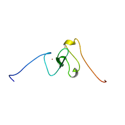
2Z0V
 | | Crystal structure of BAR domain of Endophilin-III | | 分子名称: | SH3-containing GRB2-like protein 3 | | 著者 | Murayama, K, Kato-Murayama, M, Terada, T, Shirouzu, M, Yokoyama, S, RIKEN Structural Genomics/Proteomics Initiative (RSGI) | | 登録日 | 2007-05-07 | | 公開日 | 2008-05-13 | | 最終更新日 | 2024-10-23 | | 実験手法 | X-RAY DIFFRACTION (2.49 Å) | | 主引用文献 | Crystal structure of BAR domain of Endophilin-III
To be Published
|
|
2YY1
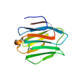 | | Crystal structure of N-terminal domain of human galectin-9 containing L-acetyllactosamine | | 分子名称: | Galectin-9, beta-D-galactopyranose-(1-4)-2-acetamido-2-deoxy-alpha-D-glucopyranose | | 著者 | Kishishita, S, Nishino, A, Murayama, K, Terada, T, Shirouzu, M, Yokoyama, S, RIKEN Structural Genomics/Proteomics Initiative (RSGI) | | 登録日 | 2007-04-27 | | 公開日 | 2008-04-29 | | 最終更新日 | 2024-03-13 | | 実験手法 | X-RAY DIFFRACTION (2.17 Å) | | 主引用文献 | Crystal structure of N-terminal domain of human galectin-9 containing L-acetyllactosamine
To be Published
|
|


