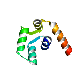
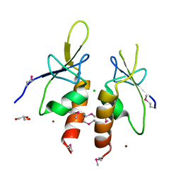
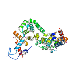
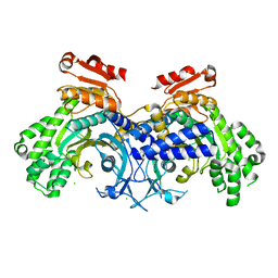
7YXP
 
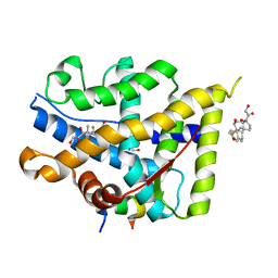 | | Crystal structure of WT AncGR2-LBD WT bound to dexamethasone and SHP coregulator fragment | | 分子名称: | 2-AMINO-2-HYDROXYMETHYL-PROPANE-1,3-DIOL, Ancestral Glucocorticoid Receptor2, DEXAMETHASONE, ... | | 著者 | Jimenez-Panizo, A, Estebanez-Perpina, E, Fuentes-Prior, P. | | 登録日 | 2022-02-16 | | 公開日 | 2022-12-07 | | 最終更新日 | 2024-01-31 | | 実験手法 | X-RAY DIFFRACTION (3.36 Å) | | 主引用文献 | The multivalency of the glucocorticoid receptor ligand-binding domain explains its manifold physiological activities.
Nucleic Acids Res., 50, 2022
|
|
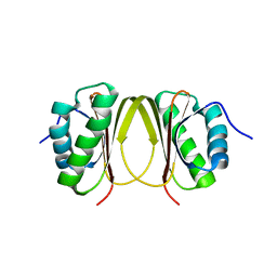
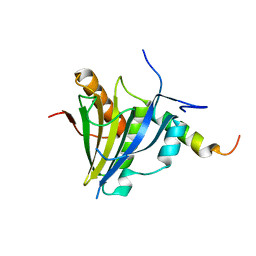
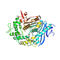
5HWT
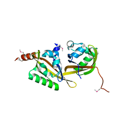 | | Crystal structure of apo-PAS1 | | 分子名称: | Sensor histidine kinase TodS | | 著者 | Hwang, J, Koh, S. | | 登録日 | 2016-01-29 | | 公開日 | 2016-03-02 | | 最終更新日 | 2017-12-06 | | 実験手法 | X-RAY DIFFRACTION (1.7 Å) | | 主引用文献 | Molecular Insights into Toluene Sensing in the TodS/TodT Signal Transduction System.
J. Biol. Chem., 291, 2016
|
|
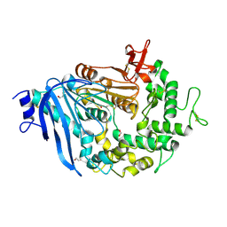
5HWW
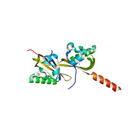 | | Crystal structure of PAS1 complexed with 1,2,4-TMB | | 分子名称: | 1,2,4-trimethylbenzene, Sensor histidine kinase TodS | | 著者 | Hwang, J, Koh, S. | | 登録日 | 2016-01-29 | | 公開日 | 2016-03-02 | | 最終更新日 | 2024-03-20 | | 実験手法 | X-RAY DIFFRACTION (2 Å) | | 主引用文献 | Molecular Insights into Toluene Sensing in the TodS/TodT Signal Transduction System.
J. Biol. Chem., 291, 2016
|
|
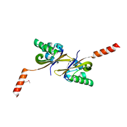
5HWV
 | |
5X2E
 | |
5X2D
 | |
5H3H
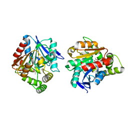 | | Esterase (EaEST) from Exiguobacterium antarcticum | | 分子名称: | Abhydrolase domain-containing protein, ETHANEPEROXOIC ACID | | 著者 | Lee, J.H, Lee, C.W. | | 登録日 | 2016-10-24 | | 公開日 | 2017-01-11 | | 最終更新日 | 2024-05-29 | | 実験手法 | X-RAY DIFFRACTION (1.9 Å) | | 主引用文献 | Crystal Structure and Functional Characterization of an Esterase (EaEST) from Exiguobacterium antarcticum.
Plos One, 12, 2017
|
|
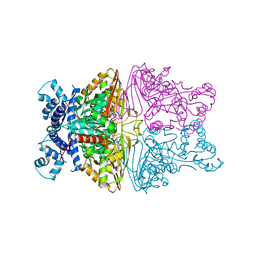
5JU6
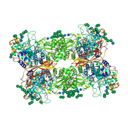 | | Structural and Functional Studies of Glycoside Hydrolase Family 3 beta-Glucosidase Cel3A from the Moderately Thermophilic Fungus Rasamsonia emersonii | | 分子名称: | 2-acetamido-2-deoxy-beta-D-glucopyranose, 2-acetamido-2-deoxy-beta-D-glucopyranose-(1-4)-2-acetamido-2-deoxy-beta-D-glucopyranose, Beta-glucosidase, ... | | 著者 | Gudmundsson, M, Sandgren, M, Karkehabadi, S. | | 登録日 | 2016-05-10 | | 公開日 | 2016-07-13 | | 最終更新日 | 2024-01-10 | | 実験手法 | X-RAY DIFFRACTION (2.2 Å) | | 主引用文献 | Structural and functional studies of the glycoside hydrolase family 3 beta-glucosidase Cel3A from the moderately thermophilic fungus Rasamsonia emersonii.
Acta Crystallogr D Struct Biol, 72, 2016
|
|
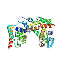
4ZYA
 | | The N-terminal extension domain of human asparaginyl-tRNA synthetase | | 分子名称: | Asparagine--tRNA ligase, cytoplasmic, CHLORIDE ION, ... | | 著者 | Park, J.S, Park, M.C, Goughnour, P, Kim, H.S, Kim, S.J, Kim, H.J, Kim, S.H, Han, B.W. | | 登録日 | 2015-05-21 | | 公開日 | 2016-05-25 | | 最終更新日 | 2019-04-24 | | 実験手法 | X-RAY DIFFRACTION (1.65 Å) | | 主引用文献 | Unique N-terminal extension domain of human asparaginyl-tRNA synthetase elicits CCR3-mediated chemokine activity.
Int. J. Biol. Macromol., 120, 2018
|
|
5HZU
 | | Crystal structure of Dronpa-Ni2+ | | 分子名称: | Fluorescent protein Dronpa, NICKEL (II) ION | | 著者 | Hwang, K.Y, Nam, K.H. | | 登録日 | 2016-02-03 | | 公開日 | 2017-03-15 | | 最終更新日 | 2023-11-15 | | 実験手法 | X-RAY DIFFRACTION (1.89 Å) | | 主引用文献 | Crystal structures of Dronpa complexed with quenchable metal ions provide insight into metal biosensor development
FEBS Lett., 590, 2016
|
|
5HZS
 | | Crystal structure of Dronpa-Co2+ | | 分子名称: | COBALT (II) ION, Fluorescent protein Dronpa | | 著者 | Hwang, K.Y, Nam, K.H. | | 登録日 | 2016-02-03 | | 公開日 | 2017-03-15 | | 最終更新日 | 2023-11-15 | | 実験手法 | X-RAY DIFFRACTION (2.17 Å) | | 主引用文献 | Crystal structures of Dronpa complexed with quenchable metal ions provide insight into metal biosensor development
FEBS Lett., 590, 2016
|
|
5HZT
 | | Crystal structure of Dronpa-Cu2+ | | 分子名称: | COPPER (II) ION, Fluorescent protein Dronpa | | 著者 | Hwang, K.Y, Nam, K.H. | | 登録日 | 2016-02-03 | | 公開日 | 2017-03-15 | | 最終更新日 | 2023-11-15 | | 実験手法 | X-RAY DIFFRACTION (2.84 Å) | | 主引用文献 | Crystal structures of Dronpa complexed with quenchable metal ions provide insight into metal biosensor development
FEBS Lett., 590, 2016
|
|
7D01
 | |
7D02
 | |
7D04
 | |
7D05
 | |
5XLN
 | |
8JB1
 | |
5X1E
 | |
5X1U
 | |
6JTT
 | | MHETase in complex with BHET | | 分子名称: | 4-(2-hydroxyethyloxycarbonyl)benzoic acid, CALCIUM ION, Mono(2-hydroxyethyl) terephthalate hydrolase, ... | | 著者 | Sagong, H.-Y, Seo, H, Kim, K.-J. | | 登録日 | 2019-04-12 | | 公開日 | 2020-04-15 | | 最終更新日 | 2020-10-28 | | 実験手法 | X-RAY DIFFRACTION (2.51 Å) | | 主引用文献 | Decomposition of PET film by MHETase using Exo-PETase function
Acs Catalysis, 10, 2020
|
|
6JTU
 | | Crystal structure of MHETase from Ideonella sakaiensis | | 分子名称: | 1,2-ETHANEDIOL, ACETATE ION, CALCIUM ION, ... | | 著者 | Sagong, H.-Y, Seo, H, Kim, K.-J. | | 登録日 | 2019-04-12 | | 公開日 | 2020-04-15 | | 最終更新日 | 2020-10-28 | | 実験手法 | X-RAY DIFFRACTION (2.1 Å) | | 主引用文献 | Decomposition of PET film by MHETase using Exo-PETase function
Acs Catalysis, 10, 2020
|
|
5X1H
 | |
6O76
 | |
7WRS
 | |


