2G4Z
 
 | | anomalous substructure of thermolysin | | 分子名称: | CALCIUM ION, CHLORIDE ION, SULFATE ION, ... | | 著者 | Mueller-Dieckmann, C, Weiss, M.S. | | 登録日 | 2006-02-22 | | 公開日 | 2007-02-20 | | 最終更新日 | 2024-02-14 | | 実験手法 | X-RAY DIFFRACTION (1.98 Å) | | 主引用文献 | On the routine use of soft X-rays in macromolecular crystallography. Part IV. Efficient determination of anomalous substructures in biomacromolecules using longer X-ray wavelengths.
Acta Crystallogr.,Sect.D, 63, 2007
|
|
2G4K
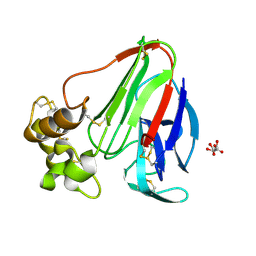
 | | Anomalous substructure of human ADP-ribosylhydrolase 3 | | 分子名称: | ADP-ribosylhydrolase 3, CHLORIDE ION, MAGNESIUM ION | | 著者 | Mueller-Dieckmann, C, Weiss, M.S. | | 登録日 | 2006-02-22 | | 公開日 | 2007-02-20 | | 最終更新日 | 2024-02-14 | | 実験手法 | X-RAY DIFFRACTION (1.82 Å) | | 主引用文献 | On the routine use of soft X-rays in macromolecular crystallography. Part IV. Efficient determination of anomalous substructures in biomacromolecules using longer X-ray wavelengths.
Acta Crystallogr.,Sect.D, 63, 2007
|
|
2G4P
 | | Anomalous substructure of lysozyme at pH 4.5 | | 分子名称: | CHLORIDE ION, Lysozyme C | | 著者 | Mueller-Dieckmann, C, Weiss, M.S. | | 登録日 | 2006-02-22 | | 公開日 | 2007-02-20 | | 最終更新日 | 2011-07-13 | | 実験手法 | X-RAY DIFFRACTION (1.84 Å) | | 主引用文献 | On the routine use of soft X-rays in macromolecular crystallography. Part IV. Efficient determination of anomalous substructures in biomacromolecules using longer X-ray wavelengths.
Acta Crystallogr.,Sect.D, 63, 2007
|
|
2G4T
 | |
2G4U
 | |
2G4H
 | | Anomalous substructure of apoferritin | | 分子名称: | CADMIUM ION, CHLORIDE ION, Ferritin light chain | | 著者 | Mueller-Dieckmann, C, Weiss, M.S. | | 登録日 | 2006-02-22 | | 公開日 | 2007-03-06 | | 最終更新日 | 2024-02-14 | | 実験手法 | X-RAY DIFFRACTION (2 Å) | | 主引用文献 | On the routine use of soft X-rays in macromolecular crystallography. Part IV. Efficient determination of anomalous substructures in biomacromolecules using longer X-ray wavelengths.
Acta Crystallogr.,Sect.D, 63, 2007
|
|
2G4L
 | | Anomalous substructure of hydroxynitrile lyase | | 分子名称: | (S)-acetone-cyanohydrin lyase, CHLORIDE ION, SULFATE ION | | 著者 | Mueller-Dieckmann, C, Weiss, M.S. | | 登録日 | 2006-02-22 | | 公開日 | 2007-02-20 | | 最終更新日 | 2011-07-13 | | 実験手法 | X-RAY DIFFRACTION (1.84 Å) | | 主引用文献 | On the routine use of soft X-rays in macromolecular crystallography. Part IV. Efficient determination of anomalous substructures in biomacromolecules using longer X-ray wavelengths.
Acta Crystallogr.,Sect.D, 63, 2007
|
|
2G4N
 | | Anomalous substructure of alpha-lactalbumin | | 分子名称: | Alpha-lactalbumin, CALCIUM ION, POTASSIUM ION | | 著者 | Mueller-Dieckmann, C, Weiss, M.S. | | 登録日 | 2006-02-22 | | 公開日 | 2007-02-20 | | 最終更新日 | 2011-07-13 | | 実験手法 | X-RAY DIFFRACTION (2.3 Å) | | 主引用文献 | On the routine use of soft X-rays in macromolecular crystallography. Part IV. Efficient determination of anomalous substructures in biomacromolecules using longer X-ray wavelengths.
Acta Crystallogr.,Sect.D, 63, 2007
|
|
2G4Y
 | | structure of thaumatin at 2.0 A wavelength | | 分子名称: | D(-)-TARTARIC ACID, Thaumatin-1 | | 著者 | Mueller-Dieckmann, C, Weiss, M.S. | | 登録日 | 2006-02-22 | | 公開日 | 2007-02-20 | | 最終更新日 | 2011-07-13 | | 実験手法 | X-RAY DIFFRACTION (1.98 Å) | | 主引用文献 | On the routine use of soft X-rays in macromolecular crystallography. Part IV. Efficient determination of anomalous substructures in biomacromolecules using longer X-ray wavelengths.
Acta Crystallogr.,Sect.D, 63, 2007
|
|
3BNG
 | | W. succinogenes NrfA Y218F | | 分子名称: | ACETATE ION, CALCIUM ION, Cytochrome c-552, ... | | 著者 | Lukat, P, Einsle, O. | | 登録日 | 2007-12-14 | | 公開日 | 2008-02-26 | | 最終更新日 | 2023-11-01 | | 実験手法 | X-RAY DIFFRACTION (1.5 Å) | | 主引用文献 | Binding and Reduction of Sulfite by Cytochrome c Nitrite Reductase
Biochemistry, 47, 2008
|
|
3BNJ
 | |
3BNF
 | | W. succinogenes NrfA Sulfite Complex | | 分子名称: | ACETATE ION, CALCIUM ION, Cytochrome c-552, ... | | 著者 | Lukat, P, Einsle, O. | | 登録日 | 2007-12-14 | | 公開日 | 2008-02-26 | | 最終更新日 | 2023-11-01 | | 実験手法 | X-RAY DIFFRACTION (1.7 Å) | | 主引用文献 | Binding and Reduction of Sulfite by Cytochrome c Nitrite Reductase
Biochemistry, 47, 2008
|
|
3BNH
 | |
1NZR
 | | CRYSTAL STRUCTURE OF THE AZURIN MUTANT NICKEL-TRP48MET FROM PSEUDOMONAS AERUGINOSA AT 2.2 ANGSTROMS RESOLUTION | | 分子名称: | AZURIN, NICKEL (II) ION, NITRATE ION | | 著者 | Tsai, L.-C, Sjolin, L, Langer, V, Bonander, N, Karlsson, B.G, Vanngard, T, Hammann, C, Nar, H. | | 登録日 | 1994-12-09 | | 公開日 | 1995-02-27 | | 最終更新日 | 2024-06-05 | | 実験手法 | X-RAY DIFFRACTION (2.2 Å) | | 主引用文献 | Structure of the azurin mutant nickel-Trp48Met from Pseudomonas aeruginosa at 2.2 A resolution.
Acta Crystallogr.,Sect.D, 51, 1995
|
|
6V3P
 | |
2IDF
 | | P. aeruginosa azurin N42C/M64E double mutant, BMME-linked dimer | | 分子名称: | 1-[PYRROL-1-YL-2,5-DIONE-METHOXYMETHYL]-PYRROLE-2,5-DIONE, Azurin, COPPER (II) ION, ... | | 著者 | Einsle, O, de Jongh, T.E, Hoffmann, M, Cavazzini, D, Rossi, G.L, Ubbink, M, Canters, G.W. | | 登録日 | 2006-09-15 | | 公開日 | 2008-03-18 | | 最終更新日 | 2023-08-30 | | 実験手法 | X-RAY DIFFRACTION (2.25 Å) | | 主引用文献 | Electron transfer in a crosslinked protein dimer mediated by a hydrogen-bonded network across the dimer interface
To be Published
|
|
8BWW
 | |
3SI7
 | | The crystal structure of the NBD1 domain of the mouse CFTR protein, deltaF508 mutant | | 分子名称: | ACETATE ION, ADENOSINE-5'-TRIPHOSPHATE, Cystic fibrosis transmembrane conductance regulator, ... | | 著者 | Brautigam, C.A, Caspa, E, Thomas, P.J. | | 登録日 | 2011-06-17 | | 公開日 | 2012-02-01 | | 最終更新日 | 2024-02-28 | | 実験手法 | X-RAY DIFFRACTION (2.25 Å) | | 主引用文献 | Requirements for efficient correction of DeltaF508 CFTR revealed by analyses of evolved sequences
Cell(Cambridge,Mass.), 148, 2012
|
|
6R26
 | | The photosensory core module (PAS-GAF-PHY) of the bacterial phytochrome Agp1 (AtBphP1) locked in a Pr-like state | | 分子名称: | 3-[2-[(~{Z})-[12-ethyl-6-(3-hydroxy-3-oxopropyl)-13-methyl-11-oxidanylidene-4,10-diazatricyclo[8.3.0.0^{3,7}]trideca-1,3,6,12-tetraen-5-ylidene]methyl]-5-[(~{Z})-(3-ethyl-4-methyl-5-oxidanylidene-pyrrol-2-ylidene)methyl]-4-methyl-1~{H}-pyrrol-3-yl]propanoic acid, Bacteriophytochrome protein, CALCIUM ION | | 著者 | Scheerer, P, Michael, N, Lamparter, T, Krauss, N. | | 登録日 | 2019-03-15 | | 公開日 | 2020-04-01 | | 最終更新日 | 2024-01-24 | | 実験手法 | X-RAY DIFFRACTION (3.11 Å) | | 主引用文献 | Crystal structures of the photosensory core module of bacteriophytochrome Agp1 reveal pronounced structural flexibility of this protein in the red-absorbing Pr state
To Be Published
|
|
6R27
 | | Crystallographic superstructure of the photosensory core module (PAS-GAF-PHY) of the bacterial phytochrome Agp1 (AtBphP1) locked in a Pr-like state | | 分子名称: | 3-[2-[(~{Z})-[12-ethyl-6-(3-hydroxy-3-oxopropyl)-13-methyl-11-oxidanylidene-4,10-diazatricyclo[8.3.0.0^{3,7}]trideca-1,3,6,12-tetraen-5-ylidene]methyl]-5-[(~{Z})-(3-ethyl-4-methyl-5-oxidanylidene-pyrrol-2-ylidene)methyl]-4-methyl-1~{H}-pyrrol-3-yl]propanoic acid, Bacteriophytochrome protein | | 著者 | Scheerer, P, Michael, N, Lamparter, T, Krauss, N. | | 登録日 | 2019-03-15 | | 公開日 | 2020-04-01 | | 最終更新日 | 2024-01-24 | | 実験手法 | X-RAY DIFFRACTION (3.4 Å) | | 主引用文献 | Crystal structures of the photosensory core module of bacteriophytochrome Agp1 reveal pronounced structural flexibility of this protein in the red-absorbing Pr state
To Be Published
|
|
6YL4
 | | Soluble epoxide hydrolase in complex with 3-((R)-3-(1-hydroxyureido)but-1-yn-1-yl)-N-((S)-3-phenyl-3-(4-trifluoromethoxy)phenyl)propyl)benzamide | | 分子名称: | 3-[(3~{R})-3-[aminocarbonyl(oxidanyl)amino]but-1-ynyl]-~{N}-[(3~{S})-3-phenyl-3-[4-(trifluoromethyloxy)phenyl]propyl]benzamide, Bifunctional epoxide hydrolase 2 | | 著者 | Kramer, J.S, Pogoryelov, D, Hiesinger, K, Proschak, E. | | 登録日 | 2020-04-06 | | 公開日 | 2020-10-21 | | 最終更新日 | 2024-05-15 | | 実験手法 | X-RAY DIFFRACTION (2.001 Å) | | 主引用文献 | Design, Synthesis, and Structure-Activity Relationship Studies of Dual Inhibitors of Soluble Epoxide Hydrolase and 5-Lipoxygenase.
J.Med.Chem., 63, 2020
|
|
2BDG
 | | Human Kallikrein 4 complex with nickel and p-aminobenzamidine | | 分子名称: | CHLORIDE ION, Kallikrein-4, NICKEL (II) ION, ... | | 著者 | Debela, M, Bode, W, Goettig, P, Structural Proteomics in Europe (SPINE) | | 登録日 | 2005-10-20 | | 公開日 | 2006-10-03 | | 最終更新日 | 2024-04-03 | | 実験手法 | X-RAY DIFFRACTION (1.95 Å) | | 主引用文献 | Crystal structures of human tissue kallikrein 4: activity modulation by a specific zinc binding site.
J.Mol.Biol., 362, 2006
|
|
2BDH
 | | Human Kallikrein 4 complex with zinc and p-aminobenzamidine | | 分子名称: | Kallikrein-4, P-AMINO BENZAMIDINE, ZINC ION | | 著者 | Debela, M, Bode, W, Goettig, P, Structural Proteomics in Europe (SPINE) | | 登録日 | 2005-10-20 | | 公開日 | 2006-10-03 | | 最終更新日 | 2024-04-03 | | 実験手法 | X-RAY DIFFRACTION (3 Å) | | 主引用文献 | Crystal structures of human tissue kallikrein 4: activity modulation by a specific zinc binding site.
J.Mol.Biol., 362, 2006
|
|
2BDI
 | | Human Kallikrein 4 complex with cobalt and p-aminobenzamidine | | 分子名称: | COBALT (II) ION, Kallikrein-4, P-AMINO BENZAMIDINE | | 著者 | Debela, M, Bode, W, Goettig, P, Structural Proteomics in Europe (SPINE) | | 登録日 | 2005-10-20 | | 公開日 | 2006-10-03 | | 最終更新日 | 2024-04-03 | | 実験手法 | X-RAY DIFFRACTION (3 Å) | | 主引用文献 | Crystal structures of human tissue kallikrein 4: activity modulation by a specific zinc binding site.
J.Mol.Biol., 362, 2006
|
|
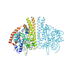
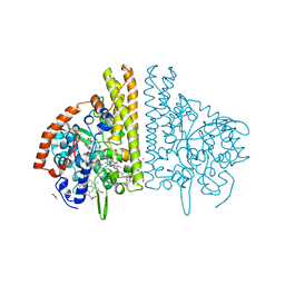
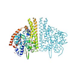
1CHM
 | | ENZYMATIC MECHANISM OF CREATINE AMIDINOHYDROLASE AS DEDUCED FROM CRYSTAL STRUCTURES | | 分子名称: | CARBAMOYL SARCOSINE, CREATINE AMIDINOHYDROLASE | | 著者 | Hoeffken, H.W, Knof, S.H, Bartlett, P.A, Huber, R, Moellering, H, Schumacher, G. | | 登録日 | 1993-07-19 | | 公開日 | 1994-04-30 | | 最終更新日 | 2024-02-07 | | 実験手法 | X-RAY DIFFRACTION (1.9 Å) | | 主引用文献 | Enzymatic mechanism of creatine amidinohydrolase as deduced from crystal structures.
J.Mol.Biol., 214, 1990
|
|


