






 9QS6
9QS6
Cryo-EM structure of the helix-stabilized MMM ubiquitin ligase complex with nanobody 270 (Composite map)
これはPDB形式変換不可エントリーです。
9QS6 の概要
| エントリーDOI | 10.2210/pdb9qs6/pdb |
| 関連するPDBエントリー | 9QRU 9QS3 9QSH |
| EMDBエントリー | 53329 |
| 分子名称 | Isoform 4 of E3 ubiquitin-protein ligase MGRN1, Modulator of smoothened protein, Nanobody 270, ... (7 entities in total) |
| 機能のキーワード | e3 ubiquitin ligase, hedgehog signaling, single-pass membrane protein, membrane protein complex, smoothened, tetraspanin, cell surface receptor, primary cilium, morphogen, signal transduction, human, carpenter syndrome, cancer, nanobody, palmitoylation, gdn, membrane protein |
| 由来する生物種 | Homo sapiens (human) 詳細 |
| タンパク質・核酸の鎖数 | 4 |
| 化学式量合計 | 147580.73 |
| 構造登録者 | Williams, C.,Carrique, L.,Pardon, E.,Nocka, L.M.,Hedger, G.,Pusapati, G.V.,Parashara, P.,Latorraca, N.R.,Sarkar, P.,Lartey, D.,Gao, L.,Milenkovic, L.,Steyaert, J.,Bazan, F.,Rouse, S.,Marqusee, S.,Kong, J.H.,Rohatgi, R.,Siebold, C. (登録日: 2025-04-04, 公開日: 2026-05-27, 最終更新日: 2026-06-17) |
| 主引用文献 | Williams, C.,Nocka, L.M.,Hedger, G.,Parashara, P.,Pardon, E.,Latorraca, N.R.,Pusapati, G.V.,Sarkar, P.,Lartey, D.,Gao, L.,Milenkovic, L.,Chalk, R.,Steyaert, J.,Marqusee, S.,Carrique, L.,Bazan, J.F.,Rouse, S.L.,Kong, J.H.,Siebold, C.,Rohatgi, R. Design principles of a membrane-spanning ubiquitin ligase. Mol.Cell, 86:2207-, 2026 Cited by PubMed Abstract: Receptor-type E3 ubiquitin ligases enable extracellular signals to control ubiquitylation in the cytoplasm, playing widespread roles in development, metabolism, and immunity. Using cryoelectron microscopy, integrated with biophysical and functional studies, we visualized a human E3 complex composed of two transmembrane proteins, MEGF8 and MOSMO, and the intracellular RING-family protein MGRN1. This MEGF8-MOSMO-MGRN1 (MMM) complex attenuates Hedgehog signaling by ubiquitylating Smoothened (SMO), a G-protein-coupled receptor (GPCR) that transduces morphogen signals. A long helix in the MMM complex engages SMO using an intramembrane degron and extends into the cytoplasm to suspend an activated and precisely oriented RING domain below the plasma membrane. This architecture enables ubiquitylation of the cytoplasmic surface of SMO, reducing SMO abundance at primary cilia. Our structure provides insights into MEGF8 mutations, which cause multi-organ birth defects, and defines a paradigm for how transmembrane E3 ligases control the cell surface abundance of GPCRs and other signaling receptors. PubMed: 42190653DOI: 10.1016/j.molcel.2026.05.001 主引用文献が同じPDBエントリー |
| 実験手法 | ELECTRON MICROSCOPY (3.3 Å) |
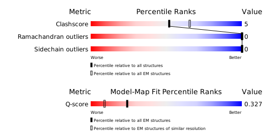
構造検証レポート
検証レポート(詳細版) をダウンロード
をダウンロード


