






 9O7T
9O7T
S. aureus YhaM D193A, 4 N-terminal domains, 2 RNA substrates
9O7T の概要
| エントリーDOI | 10.2210/pdb9o7t/pdb |
| EMDBエントリー | 70208 |
| 分子名称 | Cmp-binding-factor 1, RNA, MAGNESIUM ION, ... (4 entities in total) |
| 機能のキーワード | exonuclease, translation, rna, rna binding protein, rna binding protein-rna complex, rna binding protein/rna |
| 由来する生物種 | Staphylococcus aureus 詳細 |
| タンパク質・核酸の鎖数 | 8 |
| 化学式量合計 | 231828.20 |
| 構造登録者 | |
| 主引用文献 | Mattingly, J.M.,Liponska, A.,Tanquary, J.,Yap, M.F.,Dunham, C.M. Structural insights into RNA recognition by the Staphylococcus aureus exoribonuclease YhaM. Proc.Natl.Acad.Sci.USA, 123:e2600028123-e2600028123, 2026 Cited by PubMed Abstract: Bacterial ribonucleases regulate gene expression in response to environmental stress and host interactions. In the hibernation-promoting factor (Hpf) induces the formation of RNase R-resistant 100S ribosomes. We previously showed that the 3'-5' exoribonuclease YhaM cleaves the transcript, reducing Hpf synthesis and leading to ribosome degradation. No structure of any YhaM homolog bound to RNA is available, and biological investigations of YhaM remain limited. Here, we find that deletion of attenuates virulence in a infection model. We further determined electron cryomicroscopy structures of YhaM-RNA complexes. YhaM adopts a hexameric complex arranged in a ring, with its N-terminal oligonucleotide/oligosaccharide-binding (OB) domains positioned on both sides of the ring while the catalytic histidine/aspartate-rich (HD) domain active sites are buried within the interior. The OB-1'' domains recognize the hairpin by the formation of complementary minor groove interactions. RNA binding by two YhaM OB domains is mediated through engagement of both the backbones and nucleobases of the RNA substrate, where stacking of aromatic residues and nucleobases likely contributes to substrate recognition. Structures of YhaM bound to a single-stranded RNA reveal how the 3' ends of two RNAs are positioned within the HD domain poised for catalysis. Although six YhaM active sites are present, only two engage in RNA cleavage and further point to the importance of the remaining YhaM monomers as structural scaffolds for guiding RNA to the active site. In summary, these findings provide insights into the unique assembly of an understudied bacterial RNase. PubMed: 42301784DOI: 10.1073/pnas.2600028123 主引用文献が同じPDBエントリー |
| 実験手法 | ELECTRON MICROSCOPY (3.26 Å) |
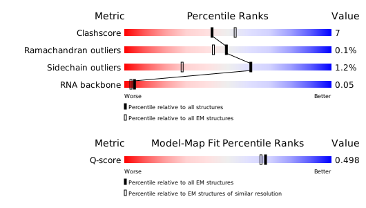
構造検証レポート
検証レポート(詳細版) をダウンロード
をダウンロード


