






 3EIL
3EIL
Structure of B-DNA d(CGTTAATTAACG)2 in the presence of Manganese
3EIL の概要
| エントリーDOI | 10.2210/pdb3eil/pdb |
| 分子名称 | 5'-D(*DCP*DGP*DTP*DTP*DAP*DAP*DTP*DTP*DAP*DAP*DCP*DG)-3', MANGANESE (II) ION (3 entities in total) |
| 機能のキーワード | b-dna, manganese, dna |
| タンパク質・核酸の鎖数 | 6 |
| 化学式量合計 | 22353.06 |
| 構造登録者 | Millonig, H.,Pous, J.,Campos, L.J.,Subirana, J.A. (登録日: 2008-09-16, 公開日: 2009-06-02, 最終更新日: 2023-08-30) |
| 主引用文献 | Millonig, H.,Pous, J.,Gouyette, C.,Subirana, J.A.,Campos, J.L. The interaction of manganese ions with DNA J.Inorg.Biochem., 103:876-880, 2009 Cited by PubMed Abstract: We present the structure of the duplex formed by a fragment of auto-complementary DNA with the sequence d(CGTTAATTAACG). The structure was determined by X-ray crystallography. Up to date it is the first structure presenting the interaction between a DNA oligonucleotide and manganese ions. The presence of Mn2+ creates bonds among the N7 atom of guanines and phosphates. These bonds stabilize and determine the crystallographic network in a P3(2) space group, unusual in oligonucleotide crystals. The crystal structure observed is compared with those found in the presence of Mg2+, Ca2+ and Ni2+, which show different kinds of interactions. The double helices show end-to-end interactions, in a manner that the terminal guanines interact with the minor groove of the neighboring duplex, while the terminal cytosines are disordered. We have chosen this sequence since it contains a TTAA repeat. Such repeats are very rare in all genomes. We suggest that this sequence may be very susceptible to the formation of closely spaced thymine dimers. PubMed: 19375803DOI: 10.1016/j.jinorgbio.2009.03.004 主引用文献が同じPDBエントリー |
| 実験手法 | X-RAY DIFFRACTION (2.6 Å) |
構造検証レポート
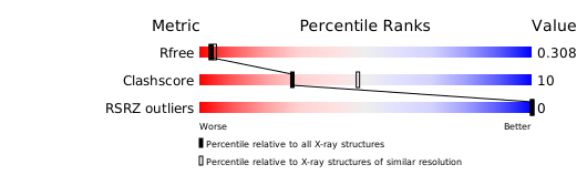
検証レポート(詳細版) をダウンロード
をダウンロード


