






 21EE
21EE
Crystal structure of human PHF20L1(1-80) in complex with H3K36me
21EE の概要
| エントリーDOI | 10.2210/pdb21ee/pdb |
| 分子名称 | PHD finger protein 20-like protein 1, Histone H3.1t (3 entities in total) |
| 機能のキーワード | tudor domain, histone binding, epigenetic reader, methyl lysine recognition, protein protein interaction, chromatin regulation, nuclear protein, structural biology, protein binding |
| 由来する生物種 | Homo sapiens (human) 詳細 |
| タンパク質・核酸の鎖数 | 2 |
| 化学式量合計 | 11511.18 |
| 構造登録者 | |
| 主引用文献 | Huang, X.,Xiao, Q.,Liu, X.,Shang, X.,Wang, Z.,Hu, H.,Zhou, Y.,Huang, Q.,Jiang, T.,Qin, S.,Huang, Y.,Li, J.B.,Liu, Y. A revised model for PHF20L1 Tudor function: DNA binding overrides methylation selectivity on nucleosomes. J.Biol.Chem., 302:113181-113181, 2026 Cited by PubMed Abstract: Plant homeodomain finger protein 20-like protein 1 (PHF20L1) is a methyl-lysine reader that regulates chromatin remodeling and transcription via its tandem Tudor and PHD finger domains. Here, we characterize the selectivity of these Tudor domains using biophysical and structural approaches. Quantitative fluorescence polarization (FP) and isothermal titration calorimetry (ITC) reveal that Tudor1 binds to both mono- and di-methylated H3K36 and H4K20, with a modest preference for H3K36me1, whereas Tudor2 is highly specific for H4K20me2. Their tandem arrangement enhances affinity for methylated H3K36 through cooperative binding. The crystal structure of Tudor1 in complex with H3K36me1 reveals the basis for its preference: a deep, narrow aromatic cage formed by Y24, Y29, F47, W50, and Y54 accommodates the mono-methylammonium group, while D23 contributes a critical hydrogen bond that stabilizes the interaction. Mutations in this cage abolish H3K36me1 binding, and mutation of corresponding cage-forming residues (W97A/Y103A, based on our solved apo Tudor2 structure) disrupts Tudor2 recognition of H4K20me2. Structural comparison across Tudor family readers for H3K36me and H4K20me further indicates that the number and orientation of acidic residues within the otherwise hydrophobic aromatic cages fine-tune selectivity among methyl-lysine states. Unexpectedly, nucleosome-binding assays reveal that the tandem Tudor domains bind to DNA with high affinity and lose methylation selectivity, contrasting sharply with the peptide-level results. Thus, our findings revise the mechanistic framework for PHF20L1 chromatin engagement: from a selective methyl-lysine reader to a high-affinity DNA-binding module on nucleosomes. PubMed: 42173250DOI: 10.1016/j.jbc.2026.113181 主引用文献が同じPDBエントリー |
| 実験手法 | X-RAY DIFFRACTION (1.34 Å) |
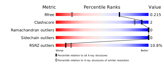
構造検証レポート
検証レポート(詳細版) をダウンロード
をダウンロード


