






 13EU
13EU
Cryo-EM structure of HAdV-C6 hexon trimer in complex with human coagulation factor X (FX)
13EU の概要
| エントリーDOI | 10.2210/pdb13eu/pdb |
| EMDBエントリー | 77027 |
| 分子名称 | Hexon protein, Coagulation factor X, CALCIUM ION (3 entities in total) |
| 機能のキーワード | adenovirus, hexon, coagulation factor x, virus-host interaction, cryo-em, virus capsid, viral protein |
| 由来する生物種 | Human adenovirus 6 詳細 |
| タンパク質・核酸の鎖数 | 4 |
| 化学式量合計 | 373746.64 |
| 構造登録者 | |
| 主引用文献 | Ma, O.X.,Lu, S.C.,Mudrick, H.E.,Barry, M.E.,French, J.B.,Barry, M.A.,Reddy, V.S. Structural requirements of blood factors binding to soluble hexon trimers with implications for adenovirus cell targeting and immune evasion. Plos Pathog., 22:e1014389-e1014389, 2026 Cited by PubMed Abstract: Human adenovirus serotype 5 (HAdV-C5) is widely used as a gene delivery vector in both experimental and clinical settings. Upon intravenous administration, HAdV-C5 exhibits strong liver tropism, largely mediated by interactions between its major capsid protein, hexon (Hx), and coagulation factor X (FX). In contrast, the closely related species C adenovirus 6 (HAdV-C6) also targets the liver but shows reduced dependency on coagulation factors, whereas species D adenovirus 26 (HAdV-D26) does not bind coagulation factors altogether. To define the structural basis of this serotype-specific host factor recognition, we determined high-resolution cryo-electron microscopy structures of isolated hexon trimers from HAdV-C5 and HAdV-C6 in complex with coagulation factors FX and prothrombin (factor II, FII). The resulting atomic models reveal conserved binding interfaces involving the γ-carboxyglutamic acid (Gla) domains of both coagulation factors and the hypervariable regions HVR5 and HVR7 lining the surface cavities of HAdV-C5 and HAdV-C6 hexons. Structures of hexon complexes formed by co-incubation with both FX and FII further reveal serotype-specific binding preferences under physiologically relevant conditions, showing that HAdV-C5 hexon preferentially engages FX, whereas HAdV-C6 hexon favors FII. By contrast, HAdV-D26 hexon does not bind either factor, likely due to an insertion constrained by proline residues in the HVR5 loop that restricts the factor access to the hexon cavity. Together, these findings provide a detailed structural framework for adenovirus-coagulation factor interactions and support the rational engineering of adenovirus vectors with improved targeting and safety profiles. PubMed: 42441716DOI: 10.1371/journal.ppat.1014389 主引用文献が同じPDBエントリー |
| 実験手法 | ELECTRON MICROSCOPY (3.26 Å) |
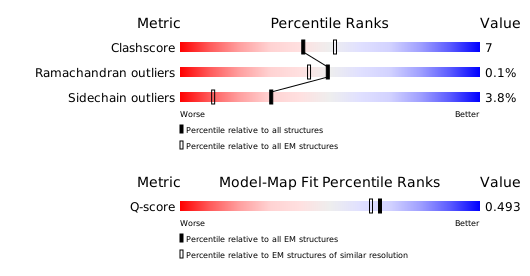
構造検証レポート
検証レポート(詳細版) をダウンロード
をダウンロード


