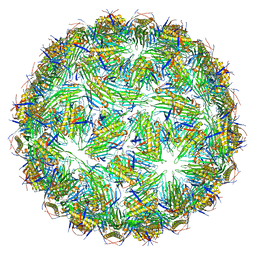
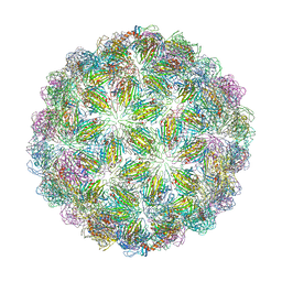
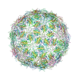
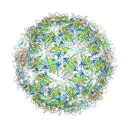
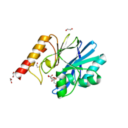
6YFC
 
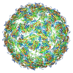 | | Virus-like particle of bacteriophage AVE019 | | 分子名称: | CALCIUM ION, coat protein | | 著者 | Rumnieks, J, Kalnins, G, Sisovs, M, Lieknina, I, Tars, K. | | 登録日 | 2020-03-26 | | 公開日 | 2020-09-02 | | 最終更新日 | 2024-01-24 | | 実験手法 | X-RAY DIFFRACTION (3.246 Å) | | 主引用文献 | Three-dimensional structure of 22 uncultured ssRNA bacteriophages: Flexibility of the coat protein fold and variations in particle shapes.
Sci Adv, 6, 2020
|
|
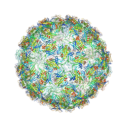
6YFD
 | | Virus-like particle of Beihai levi-like virus 14 | | 分子名称: | coat protein | | 著者 | Rumnieks, J, Kalnins, G, Sisovs, M, Lieknina, I, Tars, K. | | 登録日 | 2020-03-26 | | 公開日 | 2020-09-02 | | 最終更新日 | 2024-01-24 | | 実験手法 | X-RAY DIFFRACTION (3.3 Å) | | 主引用文献 | Three-dimensional structure of 22 uncultured ssRNA bacteriophages: Flexibility of the coat protein fold and variations in particle shapes.
Sci Adv, 6, 2020
|
|
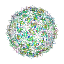
6YFE
 | | Virus-like particle of Beihai levi-like virus 19 | | 分子名称: | CALCIUM ION, coat protein | | 著者 | Rumnieks, J, Kalnins, G, Sisovs, M, Lieknina, I, Tars, K. | | 登録日 | 2020-03-26 | | 公開日 | 2020-09-02 | | 最終更新日 | 2024-01-24 | | 実験手法 | X-RAY DIFFRACTION (3.794 Å) | | 主引用文献 | Three-dimensional structure of 22 uncultured ssRNA bacteriophages: Flexibility of the coat protein fold and variations in particle shapes.
Sci Adv, 6, 2020
|
|
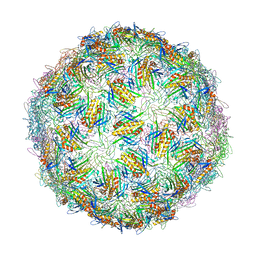
6YFF
 | | Virus-like particle of Beihai levi-like virus 21 | | 分子名称: | coat protein | | 著者 | Rumnieks, J, Kalnins, G, Sisovs, M, Lieknina, I, Tars, K. | | 登録日 | 2020-03-26 | | 公開日 | 2020-09-02 | | 最終更新日 | 2024-01-24 | | 実験手法 | X-RAY DIFFRACTION (3.089 Å) | | 主引用文献 | Three-dimensional structure of 22 uncultured ssRNA bacteriophages: Flexibility of the coat protein fold and variations in particle shapes.
Sci Adv, 6, 2020
|
|
6YFG
 | | Virus-like particle of Beihai levi-like virus 32 | | 分子名称: | CALCIUM ION, coat protein | | 著者 | Rumnieks, J, Kalnins, G, Sisovs, M, Lieknina, I, Tars, K. | | 登録日 | 2020-03-26 | | 公開日 | 2020-09-02 | | 最終更新日 | 2024-01-24 | | 実験手法 | X-RAY DIFFRACTION (3.897 Å) | | 主引用文献 | Three-dimensional structure of 22 uncultured ssRNA bacteriophages: Flexibility of the coat protein fold and variations in particle shapes.
Sci Adv, 6, 2020
|
|
6YFH
 | | Virus-like particle of bacteriophage EMS014 | | 分子名称: | coat protein | | 著者 | Rumnieks, J, Kalnins, G, Sisovs, M, Lieknina, I, Tars, K. | | 登録日 | 2020-03-26 | | 公開日 | 2020-09-02 | | 最終更新日 | 2024-01-24 | | 実験手法 | X-RAY DIFFRACTION (3.893 Å) | | 主引用文献 | Three-dimensional structure of 22 uncultured ssRNA bacteriophages: Flexibility of the coat protein fold and variations in particle shapes.
Sci Adv, 6, 2020
|
|
6YFJ
 | | Virus-like particle of bacteriophage ESE001 | | 分子名称: | coat protein | | 著者 | Rumnieks, J, Kalnins, G, Sisovs, M, Lieknina, I, Tars, K. | | 登録日 | 2020-03-26 | | 公開日 | 2020-09-02 | | 最終更新日 | 2024-11-13 | | 実験手法 | X-RAY DIFFRACTION (3.233 Å) | | 主引用文献 | Three-dimensional structure of 22 uncultured ssRNA bacteriophages: Flexibility of the coat protein fold and variations in particle shapes.
Sci Adv, 6, 2020
|
|
6YFK
 | | Virus-like particle of bacteriophage ESE007 | | 分子名称: | CALCIUM ION, coat protein | | 著者 | Rumnieks, J, Kalnins, G, Sisovs, M, Lieknina, I, Tars, K. | | 登録日 | 2020-03-26 | | 公開日 | 2020-09-02 | | 最終更新日 | 2024-01-24 | | 実験手法 | X-RAY DIFFRACTION (3.7 Å) | | 主引用文献 | Three-dimensional structure of 22 uncultured ssRNA bacteriophages: Flexibility of the coat protein fold and variations in particle shapes.
Sci Adv, 6, 2020
|
|
6YFL
 | | Virus-like particle of bacteriophage ESE020 | | 分子名称: | coat protein | | 著者 | Rumnieks, J, Kalnins, G, Sisovs, M, Lieknina, I, Tars, K. | | 登録日 | 2020-03-26 | | 公開日 | 2020-09-02 | | 最終更新日 | 2024-01-24 | | 実験手法 | X-RAY DIFFRACTION (3.3 Å) | | 主引用文献 | Three-dimensional structure of 22 uncultured ssRNA bacteriophages: Flexibility of the coat protein fold and variations in particle shapes.
Sci Adv, 6, 2020
|
|
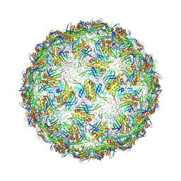
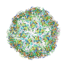
6YFM
 | | Virus-like particle of bacteriophage ESE021 | | 分子名称: | ZINC ION, coat protein | | 著者 | Rumnieks, J, Kalnins, G, Sisovs, M, Lieknina, I, Tars, K. | | 登録日 | 2020-03-26 | | 公開日 | 2020-09-02 | | 最終更新日 | 2024-01-24 | | 実験手法 | X-RAY DIFFRACTION (2.762 Å) | | 主引用文献 | Three-dimensional structure of 22 uncultured ssRNA bacteriophages: Flexibility of the coat protein fold and variations in particle shapes.
Sci Adv, 6, 2020
|
|
6YFN
 | | Virus-like particle of bacteriophage ESE058 | | 分子名称: | CALCIUM ION, coat protein | | 著者 | Rumnieks, J, Kalnins, G, Sisovs, M, Lieknina, I, Tars, K. | | 登録日 | 2020-03-26 | | 公開日 | 2020-09-02 | | 最終更新日 | 2024-01-24 | | 実験手法 | X-RAY DIFFRACTION (3.189 Å) | | 主引用文献 | Three-dimensional structure of 22 uncultured ssRNA bacteriophages: Flexibility of the coat protein fold and variations in particle shapes.
Sci Adv, 6, 2020
|
|
6YFO
 | | Virus-like particle of bacteriophage GQ-907 | | 分子名称: | coat protein | | 著者 | Rumnieks, J, Kalnins, G, Sisovs, M, Lieknina, I, Tars, K. | | 登録日 | 2020-03-26 | | 公開日 | 2020-09-02 | | 最終更新日 | 2024-01-24 | | 実験手法 | X-RAY DIFFRACTION (3.484 Å) | | 主引用文献 | Three-dimensional structure of 22 uncultured ssRNA bacteriophages: Flexibility of the coat protein fold and variations in particle shapes.
Sci Adv, 6, 2020
|
|
6YFP
 | | Virus-like particle of bacteriophage GQ-112 | | 分子名称: | coat protein | | 著者 | Rumnieks, J, Kalnins, G, Sisovs, M, Lieknina, I, Tars, K. | | 登録日 | 2020-03-26 | | 公開日 | 2020-09-02 | | 最終更新日 | 2024-01-24 | | 実験手法 | X-RAY DIFFRACTION (3.1 Å) | | 主引用文献 | Three-dimensional structure of 22 uncultured ssRNA bacteriophages: Flexibility of the coat protein fold and variations in particle shapes.
Sci Adv, 6, 2020
|
|
6YFQ
 | | Virus-like particle of bacteriophage NT-214 | | 分子名称: | coat protein | | 著者 | Rumnieks, J, Kalnins, G, Sisovs, M, Lieknina, I, Tars, K. | | 登録日 | 2020-03-26 | | 公開日 | 2020-09-02 | | 最終更新日 | 2024-10-23 | | 実験手法 | X-RAY DIFFRACTION (3.8 Å) | | 主引用文献 | Three-dimensional structure of 22 uncultured ssRNA bacteriophages: Flexibility of the coat protein fold and variations in particle shapes.
Sci Adv, 6, 2020
|
|
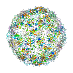
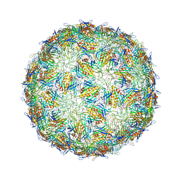
6YFR
 | | Virus-like particle of bacteriophage NT-391 | | 分子名称: | coat protein | | 著者 | Rumnieks, J, Kalnins, G, Sisovs, M, Lieknina, I, Tars, K. | | 登録日 | 2020-03-26 | | 公開日 | 2020-09-02 | | 最終更新日 | 2024-11-20 | | 実験手法 | X-RAY DIFFRACTION (2.6 Å) | | 主引用文献 | Three-dimensional structure of 22 uncultured ssRNA bacteriophages: Flexibility of the coat protein fold and variations in particle shapes.
Sci Adv, 6, 2020
|
|
6YFS
 | | Virus-like particle of bacteriophage PQ-465 | | 分子名称: | coat protein | | 著者 | Rumnieks, J, Kalnins, G, Sisovs, M, Lieknina, I, Tars, K. | | 登録日 | 2020-03-26 | | 公開日 | 2020-09-02 | | 最終更新日 | 2024-11-20 | | 実験手法 | X-RAY DIFFRACTION (3.498 Å) | | 主引用文献 | Three-dimensional structure of 22 uncultured ssRNA bacteriophages: Flexibility of the coat protein fold and variations in particle shapes.
Sci Adv, 6, 2020
|
|
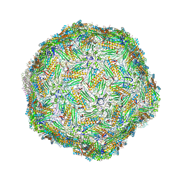
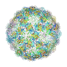
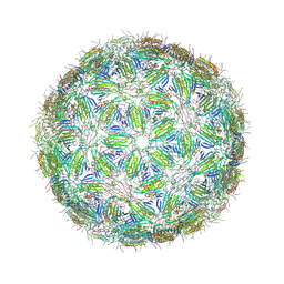
6YFT
 | | Virus-like particle of Wenzhou levi-like virus 1 | | 分子名称: | RNA, coat protein | | 著者 | Rumnieks, J, Kalnins, G, Sisovs, M, Lieknina, I, Tars, K. | | 登録日 | 2020-03-26 | | 公開日 | 2020-09-02 | | 最終更新日 | 2024-01-24 | | 実験手法 | X-RAY DIFFRACTION (3.5 Å) | | 主引用文献 | Three-dimensional structure of 22 uncultured ssRNA bacteriophages: Flexibility of the coat protein fold and variations in particle shapes.
Sci Adv, 6, 2020
|
|
6YFU
 | | Virus-like particle of Wenzhou levi-like virus 4 | | 分子名称: | coat protein | | 著者 | Rumnieks, J, Kalnins, G, Sisovs, M, Lieknina, I, Tars, K. | | 登録日 | 2020-03-26 | | 公開日 | 2020-09-02 | | 最終更新日 | 2024-01-24 | | 実験手法 | X-RAY DIFFRACTION (4.018 Å) | | 主引用文献 | Three-dimensional structure of 22 uncultured ssRNA bacteriophages: Flexibility of the coat protein fold and variations in particle shapes.
Sci Adv, 6, 2020
|
|
6YGT
 | |
6YH0
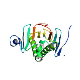
 | | Marasmius oreades agglutinin (MOA) in complex with the truncated PVPRAHS synthetic substrate | | 分子名称: | Agglutinin, CALCIUM ION, CHLORIDE ION, ... | | 著者 | Cordara, G, Manna, D, Krengel, U. | | 登録日 | 2020-03-28 | | 公開日 | 2020-07-29 | | 最終更新日 | 2024-10-23 | | 実験手法 | X-RAY DIFFRACTION (1.56 Å) | | 主引用文献 | Crystal structure of MOA in complex with a peptide fragment: A protease caught in flagranti .
Curr Res Struct Biol, 2, 2020
|
|
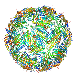
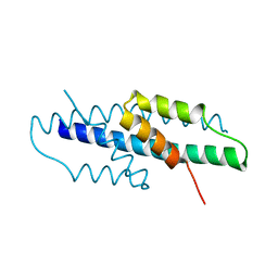
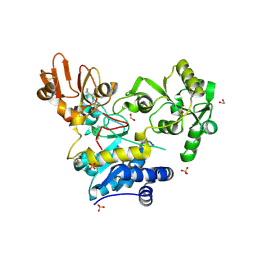
6YI0
 | | Human histidine triad nucleotide-binding protein 2 (hHINT2) refined to 1.65 A in P41212 space group | | 分子名称: | Histidine triad nucleotide-binding protein 2, mitochondrial, SODIUM ION | | 著者 | Dolot, R.D, Wlodarczyk, A, Bujacz, G.D, Nawrot, B.C. | | 登録日 | 2020-03-31 | | 公開日 | 2020-04-29 | | 最終更新日 | 2024-01-24 | | 実験手法 | X-RAY DIFFRACTION (1.65 Å) | | 主引用文献 | Biochemical, crystallographic and biophysical characterization of histidine triad nucleotide-binding protein 2 with different ligands including a non-hydrolyzable analog of Ap4A.
Biochim Biophys Acta Gen Subj, 1865, 2021
|
|
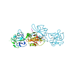
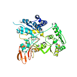
6YI4
 | | Structure of IMP-13 metallo-beta-lactamase complexed with citrate anion | | 分子名称: | 1,2-ETHANEDIOL, ACETATE ION, BETA-MERCAPTOETHANOL, ... | | 著者 | Zak, K.M, Zhou, R.X, Softley, C.A, Bostock, M.J, Sattler, M, Popowicz, G.M. | | 登録日 | 2020-03-31 | | 公開日 | 2020-04-08 | | 最終更新日 | 2024-01-24 | | 実験手法 | X-RAY DIFFRACTION (1.7 Å) | | 主引用文献 | Structure of IMP-13 metallo-beta-lactamase complexed with citrate anion
Not published
|
|
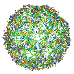
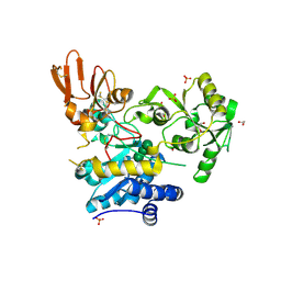
6YJQ
 | | Crystal structure of unliganded MGAT5 (alpha-1,6-mannosylglycoprotein 6-beta-N-acetylglucosaminyltransferase V) luminal domain with a Lys329-Ile345 loop truncation | | 分子名称: | 1,2-ETHANEDIOL, 2-acetamido-2-deoxy-beta-D-glucopyranose, Alpha-1,6-mannosylglycoprotein 6-beta-N-acetylglucosaminyltransferase A,Alpha-1,6-mannosylglycoprotein 6-beta-N-acetylglucosaminyltransferase A, ... | | 著者 | Wu, L, Darby, J.F, Gilio, A.K, Davies, G.J. | | 登録日 | 2020-04-04 | | 公開日 | 2020-08-05 | | 最終更新日 | 2024-11-06 | | 実験手法 | X-RAY DIFFRACTION (1.9 Å) | | 主引用文献 | Substrate Engagement and Catalytic Mechanisms of N-Acetylglucosaminyltransferase V
Acs Catalysis, 2020
|
|
6YJR
 | | Crystal structure of unliganded MGAT5 (alpha-1,6-mannosylglycoprotein 6-beta-N-acetylglucosaminyltransferase V) luminal domain. | | 分子名称: | 1,2-ETHANEDIOL, 2-acetamido-2-deoxy-beta-D-glucopyranose, Alpha-1,6-mannosylglycoprotein 6-beta-N-acetylglucosaminyltransferase A, ... | | 著者 | Wu, L, Darby, J.F, Gilio, A.K, Davies, G.J. | | 登録日 | 2020-04-04 | | 公開日 | 2020-08-05 | | 最終更新日 | 2024-11-06 | | 実験手法 | X-RAY DIFFRACTION (2.198 Å) | | 主引用文献 | Substrate Engagement and Catalytic Mechanisms of N-Acetylglucosaminyltransferase V
Acs Catalysis, 2020
|
|
6YJS
 | | Crystal structure of MGAT5 (alpha-1,6-mannosylglycoprotein 6-beta-N-acetylglucosaminyltransferase V) luminal domain with a Lys329-Ile345 loop truncation, in complex with biantennary pentasaccharide M592 | | 分子名称: | 1,2-ETHANEDIOL, 2-acetamido-2-deoxy-beta-D-glucopyranose, 2-acetamido-2-deoxy-beta-D-glucopyranose-(1-2)-alpha-D-mannopyranose-(1-3)-[2-acetamido-2-deoxy-beta-D-glucopyranose-(1-2)-alpha-D-mannopyranose-(1-6)]alpha-D-mannopyranose, ... | | 著者 | Wu, L, Darby, J.F, Gilio, A.K, Davies, G.J. | | 登録日 | 2020-04-04 | | 公開日 | 2020-08-05 | | 最終更新日 | 2024-11-20 | | 実験手法 | X-RAY DIFFRACTION (1.6 Å) | | 主引用文献 | Substrate Engagement and Catalytic Mechanisms of N-Acetylglucosaminyltransferase V
Acs Catalysis, 2020
|
|


