 Movie
Movie Controller

[English] 日本語
 Yorodumi
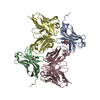
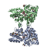
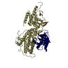
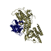
- PDB-5vrf: CryoEM Structure of the Zinc Transporter YiiP from helical crystals -
+ Open data

- Basic information
| Entry | Database: PDB / ID: 5vrf | |||||||||
|---|---|---|---|---|---|---|---|---|---|---|
| Title | CryoEM Structure of the Zinc Transporter YiiP from helical crystals | |||||||||
 Components Components | Cadmium and zinc efflux pump FieF | |||||||||
 Keywords Keywords |  MEMBRANE PROTEIN / zinc antiporter / cation diffusion facilitator / metal transport / helical crystals / Structural Genomics / PSI-Biology / Transcontinental EM Initiative for Membrane Protein Structure / TEMIMPS MEMBRANE PROTEIN / zinc antiporter / cation diffusion facilitator / metal transport / helical crystals / Structural Genomics / PSI-Biology / Transcontinental EM Initiative for Membrane Protein Structure / TEMIMPS | |||||||||
| Function / homology |  Function and homology information Function and homology informationzinc efflux active transmembrane transporter activity / cadmium ion transmembrane transporter activity / ferrous iron transmembrane transporter activity / intracellular zinc ion homeostasis / metal ion binding / plasma membraneSimilarity search - Function | |||||||||
| Biological species |  Shewanella oneidensis MR-1 (bacteria) Shewanella oneidensis MR-1 (bacteria) | |||||||||
| Method | ELECTRON MICROSCOPY / helical reconstruction / cryo EM / Resolution: 4.1 Å | |||||||||
 Authors Authors | Coudray, N. / Lopez-Redondo, M. / Zhang, Z. / Alexopoulos, J. / Stokes, D.L. / Transcontinental EM Initiative for Membrane Protein Structure (TEMIMPS) | |||||||||
| Funding support |  United States, 2items United States, 2items
| |||||||||
 Citation Citation | Journal: Proc Natl Acad Sci U S A / Year: 2018 Title: Structural basis for the alternating access mechanism of the cation diffusion facilitator YiiP. Authors: Maria Luisa Lopez-Redondo / Nicolas Coudray / Zhening Zhang / John Alexopoulos / David L Stokes / Abstract: YiiP is a dimeric antiporter from the cation diffusion facilitator family that uses the proton motive force to transport Zn across bacterial membranes. Previous work defined the atomic structure of ...YiiP is a dimeric antiporter from the cation diffusion facilitator family that uses the proton motive force to transport Zn across bacterial membranes. Previous work defined the atomic structure of an outward-facing conformation, the location of several Zn binding sites, and hydrophobic residues that appear to control access to the transport sites from the cytoplasm. A low-resolution cryo-EM structure revealed changes within the membrane domain that were associated with the alternating access mechanism for transport. In the current work, the resolution of this cryo-EM structure has been extended to 4.1 Å. Comparison with the X-ray structure defines the differences between inward-facing and outward-facing conformations at an atomic level. These differences include rocking and twisting of a four-helix bundle that harbors the Zn transport site and controls its accessibility within each monomer. As previously noted, membrane domains are closely associated in the dimeric structure from cryo-EM but dramatically splayed apart in the X-ray structure. Cysteine crosslinking was used to constrain these membrane domains and to show that this large-scale splaying was not necessary for transport activity. Furthermore, dimer stability was not compromised by mutagenesis of elements in the cytoplasmic domain, suggesting that the extensive interface between membrane domains is a strong determinant of dimerization. As with other secondary transporters, this interface could provide a stable scaffold for movements of the four-helix bundle that confers alternating access of these ions to opposite sides of the membrane. | |||||||||
| History |
|
- Structure visualization
| Movie |
Movie viewer |
|---|---|
| Structure viewer | Molecule: MolmilJmol/JSmol |
- Downloads & links
-Download
| PDBx/mmCIF format | 5vrf.cif.gz | 109.3 KB | Display | PDBx/mmCIF format |
|---|---|---|---|---|
| PDB format | pdb5vrf.ent.gz | 83.1 KB | Display | PDB format |
| PDBx/mmJSON format | 5vrf.json.gz | Tree view | PDBx/mmJSON format | |
| Others |  Other downloads Other downloads |
-Validation report
| Arichive directory | https://data.pdbj.org/pub/pdb/validation_reports/vr/5vrfftp://data.pdbj.org/pub/pdb/validation_reports/vr/5vrf | HTTPS FTP |
|---|
-Related structure data
| Related structure data |  8728MC M: map data used to model this data C: citing same article ( |
|---|---|
| Similar structure data | |
| Other databases |


-Links
 PDBj
PDBj- Assembly
| Deposited unit | 
|
|---|---|
| 1 |
|
-Components
| #1: Protein | Mass: 32485.211 Da / Num. of mol.: 2 Source method: isolated from a genetically manipulated source Source: (gene. exp.) Shewanella oneidensis MR-1 (bacteria) / Strain: MR-1 / Gene: fieF, SO_4475 / Production host: Escherichia coli BL21(DE3) (bacteria) / Strain (production host): BL21 (DE3) / References: UniProt: Q8E919#2: Chemical | ChemComp-ZN /   Mass: 65.409 Da / Num. of mol.: 8 Mass: 65.409 Da / Num. of mol.: 8Source method: isolated from a genetically manipulated source Formula: Zn |
|---|
-Experimental details
-Experiment
| Experiment | Method: ELECTRON MICROSCOPY |
|---|---|
| EM experiment | Aggregation state: HELICAL ARRAY / 3D reconstruction method: helical reconstruction |
- Sample preparation
| Component | Name: YiiP dimer in an inward-facing conformation / Type: COMPLEX / Entity ID: #1 / Source: RECOMBINANT | ||||||||||||||||||||
|---|---|---|---|---|---|---|---|---|---|---|---|---|---|---|---|---|---|---|---|---|---|
| Molecular weight | Value: 0.06 MDa / Experimental value: YES | ||||||||||||||||||||
| Source (natural) | Organism: Shewanella oneidensis MR-1 (bacteria) | ||||||||||||||||||||
| Source (recombinant) | Organism: Escherichia coli BL21(DE3) (bacteria) / Strain: BL21 (DE3) | ||||||||||||||||||||
| Buffer solution | pH: 7 / Details: Buffer was changed twice per day. | ||||||||||||||||||||
| Buffer component |
| ||||||||||||||||||||
| Specimen | Conc.: 0.6 mg/ml / Embedding applied: NO / Shadowing applied: NO / Staining applied: NO / Vitrification applied: YES Details: The protein was purified in 0.2% n-dodecyl beta-D-maltoside | ||||||||||||||||||||
| Specimen support | Grid material: COPPER / Grid mesh size: 400 divisions/in. / Grid type: EMS | ||||||||||||||||||||
| Vitrification | Instrument: HOMEMADE PLUNGER / Cryogen name: ETHANE / Humidity: 90 % |
- Electron microscopy imaging
| Experimental equipment |  Model: Titan Krios / Image courtesy: FEI Company |
|---|---|
| Microscopy | Model: FEI TITAN KRIOS |
| Electron gun | Electron source: LAB6 / Accelerating voltage: 300 kV / Illumination mode: FLOOD BEAM |
| Electron lens | Mode: BRIGHT FIELDBright-field microscopy / Nominal magnification: 22500 X / Nominal defocus max: 2500 nm / Nominal defocus min: 1200 nm / Calibrated defocus min: 1000 nm / Calibrated defocus max: 5400 nm / Cs: 2.7 mm / Alignment procedure: COMA FREE |
| Specimen holder | Cryogen: NITROGEN / Specimen holder model: FEI TITAN KRIOS AUTOGRID HOLDER |
| Image recording | Average exposure time: 10 sec. / Electron dose: 70 e/Å2 / Detector mode: COUNTING / Film or detector model: GATAN K2 SUMMIT (4k x 4k) / Num. of grids imaged: 3 / Num. of real images: 2743 |
| Image scans | Width: 3710 / Height: 3838 / Movie frames/image: 50 / Used frames/image: 2-16 |
- Processing
| Software | Name: PHENIX / Version: 1.11.1_2575: / Classification: refinement | ||||||||||||||||||||||||||||||||||||||||||||||||||||||||||||
|---|---|---|---|---|---|---|---|---|---|---|---|---|---|---|---|---|---|---|---|---|---|---|---|---|---|---|---|---|---|---|---|---|---|---|---|---|---|---|---|---|---|---|---|---|---|---|---|---|---|---|---|---|---|---|---|---|---|---|---|---|---|
| EM software |
| ||||||||||||||||||||||||||||||||||||||||||||||||||||||||||||
| CTF correction | Type: PHASE FLIPPING AND AMPLITUDE CORRECTION | ||||||||||||||||||||||||||||||||||||||||||||||||||||||||||||
| Helical symmerty | Angular rotation/subunit: -17 ° / Axial rise/subunit: 28.2 Å / Axial symmetry: D5 | ||||||||||||||||||||||||||||||||||||||||||||||||||||||||||||
| Particle selection | Num. of particles selected: 141904 Details: 2293 filaments selected with SPARX/EMAN2, then windowed into 141,904 segments (450x450 pixel overlapping segments) | ||||||||||||||||||||||||||||||||||||||||||||||||||||||||||||
| 3D reconstruction | Resolution: 4.1 Å / Resolution method: FSC 0.143 CUT-OFF / Num. of particles: 72333 Details: Reconstruction was achieved using the IHRSR method implemented in RELION 2.0. Symmetry type: HELICAL | ||||||||||||||||||||||||||||||||||||||||||||||||||||||||||||
| Atomic model building | Protocol: FLEXIBLE FIT / Space: REAL / Target criteria: cross-correlation coefficient Details: The residues were manually adjusted in COOT relying on the large side-chain residues, and the whole system was further optimized using the real_space_refine algorithm in Phenix to ensure ...Details: The residues were manually adjusted in COOT relying on the large side-chain residues, and the whole system was further optimized using the real_space_refine algorithm in Phenix to ensure proper fit. The conformation was subjected to 13 rounds of COOT/Phenix refinements. | ||||||||||||||||||||||||||||||||||||||||||||||||||||||||||||
| Atomic model building | 3D fitting-ID: 1 / Accession code: 3J1Z / Initial refinement model-ID: 1 / Pdb chain residue range: 7-288 / PDB-ID: 3J1Z / Source name: PDB / Type: experimental model
| ||||||||||||||||||||||||||||||||||||||||||||||||||||||||||||
| Refine LS restraints |
|

