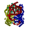
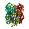
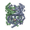
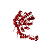
[English] 日本語
Yorodumi
- PDB-6quf: Protein crystallization by ionic liquid hydrogel support: referen... -

+
Open data


ID or keywords:

Loading...

-
Basic information

Entry
Database: PDB / ID: 6quf
TitleProtein crystallization by ionic liquid hydrogel support: reference crystal of glucose isomerase grown on standard silanized glass
ComponentsXylose isomerase
KeywordsISOMERASE / Glucose Isomerase / ionic liquid / structural comparison / hydration shell
Function / homology
Function and homology information


xylose isomerase / D-xylose metabolic process / xylose isomerase activity / magnesium ion binding / identical protein binding / cytoplasm
Similarity search - Function
Xylose isomerase, actinobacteria / Xylose isomerase / Xylose isomerase family profile. / Divalent-metal-dependent TIM barrel enzymes / Xylose isomerase-like, TIM barrel domain / Xylose isomerase-like TIM barrel / Xylose isomerase-like superfamily / TIM Barrel / Alpha-Beta Barrel / Alpha Beta
Similarity search - Domain/homology
: / Xylose isomerase
Similarity search - Component
Biological speciesStreptomyces rubiginosus (bacteria)
MethodX-RAY DIFFRACTION / SYNCHROTRON / MOLECULAR REPLACEMENT / Resolution: 1.19 Å
AuthorsBelviso, B.D. / Caliandro, R. / Caliandro, R.
CitationJournal: Crystals / Year: 2019
Title: Protein Crystallization in Ionic-Liquid Hydrogel Composite Membranes
Authors: Belviso, B.D. / Caliandro, R. / Salehi, S.M. / di Profio, G. / Caliandro, R.
History
DepositionFeb 27, 2019Deposition site: PDBE / Processing site: PDBE
Revision 1.0Mar 13, 2019Provider: repository / Type: Initial release
Revision 1.1May 22, 2019Group: Data collection / Category: reflns / Item: _reflns.B_iso_Wilson_estimate
Revision 1.2Jul 10, 2019Group: Data collection / Category: diffrn_source / Item: _diffrn_source.pdbx_synchrotron_site
Revision 1.3Sep 25, 2019Group: Data collection / Database references / Category: citation / citation_author
Item: _citation.country / _citation.journal_abbrev ..._citation.country / _citation.journal_abbrev / _citation.journal_id_CSD / _citation.journal_id_ISSN / _citation.pdbx_database_id_DOI / _citation.title / _citation.year
Revision 1.4Apr 8, 2020Group: Database references / Category: citation / Item: _citation.journal_volume / _citation.page_first
Revision 1.5Jan 24, 2024Group: Data collection / Database references / Refinement description
Category: chem_comp_atom / chem_comp_bond ...chem_comp_atom / chem_comp_bond / database_2 / pdbx_initial_refinement_model
Item: _database_2.pdbx_DOI / _database_2.pdbx_database_accession

-
Structure visualization

Structure viewerMolecule:
MolmilJmol/JSmol

Downloads & links

-
Assembly

Deposited unit
A: Xylose isomerase
hetero molecules


Theoretical massNumber of molelcules
Total (without water)43,7667
Polymers43,2831
Non-polymers4826
Water9,692538
1
A: Xylose isomerase
hetero molecules

A: Xylose isomerase
hetero molecules

A: Xylose isomerase
hetero molecules

A: Xylose isomerase
hetero molecules


Theoretical massNumber of molelcules
Total (without water)175,06228
Polymers173,1334
Non-polymers1,92924
Water724
TypeNameSymmetry operationNumber
identity operation1_555x,y,z1
crystal symmetry operation2_765-x+2,-y+1,z1
crystal symmetry operation3_757-x+2,y,-z+21
crystal symmetry operation4_567x,-y+1,-z+21
Buried area35480 Å2
ΔGint-222 kcal/mol
Surface area45440 Å2
MethodPISA
Unit cell
Length a, b, c (Å)92.522, 98.167, 101.851
Angle α, β, γ (deg.)90.00, 90.00, 90.00
Int Tables number23
Space group name H-MI222
Components on special symmetry positions
IDModelComponents
11A-903-

HOH

-
Components

#1: Protein Xylose isomerase /


Mass: 43283.297 Da / Num. of mol.: 1 / Source method: isolated from a natural source / Source: (natural) Streptomyces rubiginosus (bacteria) / References: UniProt: P24300, xylose isomerase
#2: Chemical ChemComp-MN / MANGANESE (II) ION


Mass: 54.938 Da / Num. of mol.: 2 / Source method: obtained synthetically / Formula: Mn
#3: Chemical ChemComp-GOL / GLYCEROL / GLYCERIN / PROPANE-1,2,3-TRIOL / Glycerol


Mass: 92.094 Da / Num. of mol.: 3 / Source method: obtained synthetically / Formula: C3H8O3
#4: Chemical ChemComp-SO4 / SULFATE ION / Sulfate


Mass: 96.063 Da / Num. of mol.: 1 / Source method: obtained synthetically / Formula: SO4
#5: Water ChemComp-HOH / water / Water


Mass: 18.015 Da / Num. of mol.: 538 / Source method: isolated from a natural source / Formula: H2O

-
Experimental details

-
Experiment

ExperimentMethod: X-RAY DIFFRACTION / Number of used crystals: 1

-
Sample preparation

CrystalDensity Matthews: 2.67 Å3/Da / Density % sol: 53.96 %
Crystal growTemperature: 298 K / Method: vapor diffusion / pH: 7
Details: Protein concentration 26mg/ml in HEPES 10mM pH 7 and MgCl2 1mM. reservoir: (NH4)2SO4 1.5M

-
Data collection

DiffractionMean temperature: 100 K / Serial crystal experiment: N
Diffraction sourceSource: SYNCHROTRON / Site: Diamond / Beamline: I04 / Wavelength: 0.97779 Å
DetectorType: DECTRIS EIGER X 16M / Detector: PIXEL / Date: Dec 12, 2016
RadiationMonochromator: double crystal / Protocol: SINGLE WAVELENGTH / Monochromatic (M) / Laue (L): M / Scattering type: x-ray
Radiation wavelengthWavelength: 0.97779 Å / Relative weight: 1
ReflectionResolution: 1.19→39.93 Å / Num. obs: 112879 / % possible obs: 76.7 % / Redundancy: 3.4 % / Biso Wilson estimate: 8.59 Å2 / CC1/2: 0.999 / Rmerge(I) obs: 0.042 / Rpim(I) all: 0.03 / Rrim(I) all: 0.049 / Net I/σ(I): 14.7
Reflection shellResolution: 1.19→1.22 Å / Redundancy: 1 % / Rmerge(I) obs: 0.166 / Mean I/σ(I) obs: 2.6 / Num. unique obs: 522 / CC1/2: 0.966 / Rpim(I) all: 0.166 / Rrim(I) all: 0.234 / % possible all: 4.9

-
Processing

Software
NameVersionClassification
xia2data reduction
Aimlessdata scaling
Sir2014phasing
REFMAC5.8.0238refinement
BUSTER-TNTrefinement
RefinementMethod to determine structure: MOLECULAR REPLACEMENT
Starting model: 1OAD
Resolution: 1.19→39.93 Å / Cor.coef. Fo:Fc: 0.983 / Cor.coef. Fo:Fc free: 0.978 / SU B: 0.979 / SU ML: 0.019 / Cross valid method: THROUGHOUT / ESU R: 0.032 / ESU R Free: 0.032 / Details: HYDROGENS HAVE BEEN ADDED IN THE RIDING POSITIONS
RfactorNum. reflection% reflectionSelection details
Rfree0.12943 5749 5.1 %RANDOM
Rwork0.10659 ---
obs0.10775 107045 76.54 %-
Solvent computationIon probe radii: 0.8 Å / Shrinkage radii: 0.8 Å / VDW probe radii: 1.2 Å
Displacement parametersBiso mean: 13.397 Å2
Baniso -1Baniso -2Baniso -3
1-0.01 Å20 Å20 Å2
2---0.07 Å2-0 Å2
3---0.06 Å2
Refinement stepCycle: 1 / Resolution: 1.19→39.93 Å
ProteinNucleic acidLigandSolventTotal
Num. atoms3033 0 25 538 3596
Refine LS restraints
Refine-IDTypeDev idealDev ideal targetNumber
X-RAY DIFFRACTIONr_bond_refined_d0.0040.0133285
X-RAY DIFFRACTIONr_bond_other_d0.0010.0172994
X-RAY DIFFRACTIONr_angle_refined_deg1.3471.6494466
X-RAY DIFFRACTIONr_angle_other_deg1.5171.5846926
X-RAY DIFFRACTIONr_dihedral_angle_1_deg6.6655421
X-RAY DIFFRACTIONr_dihedral_angle_2_deg30.14620.783217
X-RAY DIFFRACTIONr_dihedral_angle_3_deg11.64215529
X-RAY DIFFRACTIONr_dihedral_angle_4_deg17.6041538
X-RAY DIFFRACTIONr_chiral_restr0.0630.2398
X-RAY DIFFRACTIONr_gen_planes_refined0.0180.023838
X-RAY DIFFRACTIONr_gen_planes_other0.0080.02785
X-RAY DIFFRACTIONr_nbd_refined
X-RAY DIFFRACTIONr_nbd_other
X-RAY DIFFRACTIONr_nbtor_refined
X-RAY DIFFRACTIONr_nbtor_other
X-RAY DIFFRACTIONr_xyhbond_nbd_refined
X-RAY DIFFRACTIONr_xyhbond_nbd_other
X-RAY DIFFRACTIONr_metal_ion_refined
X-RAY DIFFRACTIONr_metal_ion_other
X-RAY DIFFRACTIONr_symmetry_vdw_refined
X-RAY DIFFRACTIONr_symmetry_vdw_other
X-RAY DIFFRACTIONr_symmetry_hbond_refined
X-RAY DIFFRACTIONr_symmetry_hbond_other
X-RAY DIFFRACTIONr_symmetry_metal_ion_refined
X-RAY DIFFRACTIONr_symmetry_metal_ion_other
X-RAY DIFFRACTIONr_mcbond_it6.9680.9621601
X-RAY DIFFRACTIONr_mcbond_other6.9660.961600
X-RAY DIFFRACTIONr_mcangle_it8.5711.4532013
X-RAY DIFFRACTIONr_mcangle_other8.5731.4542014
X-RAY DIFFRACTIONr_scbond_it10.1861.421684
X-RAY DIFFRACTIONr_scbond_other10.1891.4221685
X-RAY DIFFRACTIONr_scangle_it
X-RAY DIFFRACTIONr_scangle_other10.8451.9682441
X-RAY DIFFRACTIONr_long_range_B_refined15.2613.9553976
X-RAY DIFFRACTIONr_long_range_B_other15.34113.1013833
X-RAY DIFFRACTIONr_rigid_bond_restr26.14136276
X-RAY DIFFRACTIONr_sphericity_free
X-RAY DIFFRACTIONr_sphericity_bonded
LS refinement shellResolution: 1.191→1.222 Å / Total num. of bins used: 20
RfactorNum. reflection% reflection
Rfree0.306 26 -
Rwork0.243 500 -
obs--4.87 %

+
About Yorodumi

-
News

-
Feb 9, 2022. New format data for meta-information of EMDB entries

New format data for meta-information of EMDB entries

  • Version 3 of the EMDB header file is now the official format.
  • The previous official version 1.9 will be removed from the archive.

Related info.:EMDB header

External links:wwPDB to switch to version 3 of the EMDB data model

-
Aug 12, 2020. Covid-19 info

Covid-19 info

URL: https://pdbj.org/emnavi/covid19.php

New page: Covid-19 featured information page in EM Navigator.

Related info.:Covid-19 info / Mar 5, 2020. Novel coronavirus structure data

+
Mar 5, 2020. Novel coronavirus structure data

Novel coronavirus structure data

Related info.:Yorodumi Speices / Aug 12, 2020. Covid-19 info

External links:COVID-19 featured content - PDBj / Molecule of the Month (242):Coronavirus Proteases

+
Jan 31, 2019. EMDB accession codes are about to change! (news from PDBe EMDB page)

EMDB accession codes are about to change! (news from PDBe EMDB page)

  • The allocation of 4 digits for EMDB accession codes will soon come to an end. Whilst these codes will remain in use, new EMDB accession codes will include an additional digit and will expand incrementally as the available range of codes is exhausted. The current 4-digit format prefixed with “EMD-” (i.e. EMD-XXXX) will advance to a 5-digit format (i.e. EMD-XXXXX), and so on. It is currently estimated that the 4-digit codes will be depleted around Spring 2019, at which point the 5-digit format will come into force.
  • The EM Navigator/Yorodumi systems omit the EMD- prefix.

Related info.:Q: What is EMD? / ID/Accession-code notation in Yorodumi/EM Navigator

External links:EMDB Accession Codes are Changing Soon! / Contact to PDBj

+
Jul 12, 2017. Major update of PDB

Major update of PDB

  • wwPDB released updated PDB data conforming to the new PDBx/mmCIF dictionary.
  • This is a major update changing the version number from 4 to 5, and with Remediation, in which all the entries are updated.
  • In this update, many items about electron microscopy experimental information are reorganized (e.g. em_software).
  • Now, EM Navigator and Yorodumi are based on the updated data.

External links:wwPDB Remediation / Enriched Model Files Conforming to OneDep Data Standards Now Available in the PDB FTP Archive

-
Yorodumi

Thousand views of thousand structures

  • Yorodumi is a browser for structure data from EMDB, PDB, SASBDB, etc.
  • This page is also the successor to EM Navigator detail page, and also detail information page/front-end page for Omokage search.
  • The word "yorodu" (or yorozu) is an old Japanese word meaning "ten thousand". "mi" (miru) is to see.

Related info.:EMDB / PDB / SASBDB / Comparison of 3 databanks / Yorodumi Search / Aug 31, 2016. New EM Navigator & Yorodumi / Yorodumi Papers / Jmol/JSmol / Function and homology information / Changes in new EM Navigator and Yorodumi

Read more