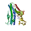
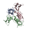
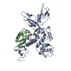
Journal: Nat Commun / Year: 2019 Title: The structure of a prokaryotic viral envelope protein expands the landscape of membrane fusion proteins. Authors: Kamel El Omari / Sai Li / Abhay Kotecha / Thomas S Walter / Eduardo A Bignon / Karl Harlos / Pentti Somerharju / Felix De Haas / Daniel K Clare / Mika Molin / Felipe Hurtado / Mengqiu Li / ...Authors: Kamel El Omari / Sai Li / Abhay Kotecha / Thomas S Walter / Eduardo A Bignon / Karl Harlos / Pentti Somerharju / Felix De Haas / Daniel K Clare / Mika Molin / Felipe Hurtado / Mengqiu Li / Jonathan M Grimes / Dennis H Bamford / Nicole D Tischler / Juha T Huiskonen / David I Stuart / Elina Roine / Abstract: Lipid membrane fusion is an essential function in many biological processes. Detailed mechanisms of membrane fusion and the protein structures involved have been mainly studied in eukaryotic systems, ...Lipid membrane fusion is an essential function in many biological processes. Detailed mechanisms of membrane fusion and the protein structures involved have been mainly studied in eukaryotic systems, whereas very little is known about membrane fusion in prokaryotes. Haloarchaeal pleomorphic viruses (HRPVs) have a membrane envelope decorated with spikes that are presumed to be responsible for host attachment and membrane fusion. Here we determine atomic structures of the ectodomains of the 57-kDa spike protein VP5 from two related HRPVs revealing a previously unreported V-shaped fold. By Volta phase plate cryo-electron tomography we show that VP5 is monomeric on the viral surface, and we establish the orientation of the molecules with respect to the viral membrane. We also show that the viral membrane fuses with the host cytoplasmic membrane in a process mediated by VP5. This sheds light on protein structures involved in prokaryotic membrane fusion.
Method to determine structure: SAD / Resolution: 2.69→82.35 Å / Cor.coef. Fo:Fc: 0.915 / Cor.coef. Fo:Fc free: 0.899 / SU R Cruickshank DPI: 0.273 / Cross valid method: THROUGHOUT / σ(F): 0 / SU R Blow DPI: 0.283 / SU Rfree Blow DPI: 0.216 / SU Rfree Cruickshank DPI: 0.213
Rfactor
Num. reflection
% reflection
Selection details
Rfree
0.233
2394
4.88 %
RANDOM
Rwork
0.218
-
-
-
obs
0.219
49062
99.9 %
-
Displacement parameters
Biso mean: 77.93 Å2
Baniso -1
Baniso -2
Baniso -3
1-
11.5442 Å2
0 Å2
0 Å2
2-
-
11.5442 Å2
0 Å2
3-
-
-
-23.0884 Å2
Refine analyze
Luzzati coordinate error obs: 0.41 Å
Refinement step
Cycle: 1 / Resolution: 2.69→82.35 Å
Protein
Nucleic acid
Ligand
Solvent
Total
Num. atoms
5493
0
25
196
5714
Refine LS restraints
Refine-ID
Type
Dev ideal
Number
Restraint function
Weight
X-RAY DIFFRACTION
t_bond_d
0.007
5588
HARMONIC
2
X-RAY DIFFRACTION
t_angle_deg
0.99
7633
HARMONIC
2
X-RAY DIFFRACTION
t_dihedral_angle_d
1875
SINUSOIDAL
2
X-RAY DIFFRACTION
t_incorr_chiral_ct
X-RAY DIFFRACTION
t_pseud_angle
X-RAY DIFFRACTION
t_trig_c_planes
X-RAY DIFFRACTION
t_gen_planes
986
HARMONIC
5
X-RAY DIFFRACTION
t_it
5588
HARMONIC
20
X-RAY DIFFRACTION
t_nbd
X-RAY DIFFRACTION
t_omega_torsion
2.16
X-RAY DIFFRACTION
t_other_torsion
15.04
X-RAY DIFFRACTION
t_improper_torsion
X-RAY DIFFRACTION
t_chiral_improper_torsion
792
SEMIHARMONIC
5
X-RAY DIFFRACTION
t_sum_occupancies
X-RAY DIFFRACTION
t_utility_distance
X-RAY DIFFRACTION
t_utility_angle
X-RAY DIFFRACTION
t_utility_torsion
X-RAY DIFFRACTION
t_ideal_dist_contact
6402
SEMIHARMONIC
4
LS refinement shell
Resolution: 2.69→2.71 Å / Total num. of bins used: 50
Rfactor
Num. reflection
% reflection
Rfree
0.2872
-
5.19 %
Rwork
0.2827
931
-
all
0.2829
982
-
obs
-
-
99.69 %
Refinement TLS params.
Method: refined / Refine-ID: X-RAY DIFFRACTION
ID
L11 (°2)
L12 (°2)
L13 (°2)
L22 (°2)
L23 (°2)
L33 (°2)
S11 (Å °)
S12 (Å °)
S13 (Å °)
S21 (Å °)
S22 (Å °)
S23 (Å °)
S31 (Å °)
S32 (Å °)
S33 (Å °)
T11 (Å2)
T12 (Å2)
T13 (Å2)
T22 (Å2)
T23 (Å2)
T33 (Å2)
Origin x (Å)
Origin y (Å)
Origin z (Å)
1
1.5037
-2.162
-0.238
4.0366
-0.3793
0.7967
-0.0132
0.0596
-0.0482
0.15
-0.1175
0.072
-0.0649
0.0233
0.1307
0.1292
0.2339
0.1195
-0.1869
0.0585
0.0304
15.9628
47.4417
206.98
2
2.581
-2.6696
-2.1301
1.7782
2.6656
2.1075
0.0221
-0.2087
-0.1333
0.004
-0.1402
-0.0567
0.1851
0.135
0.1181
0.4383
0.1175
0.2834
-0.3342
0.1062
-0.0541
7.8672
40.1848
231.806
3
1.9683
-2.8974
0.3239
5.7215
-1.5626
0.7976
-0.1064
-0.0105
-0.0675
0.2869
-0.0425
0.0029
0.0292
0.0671
0.1489
0.226
0.2135
0.0555
-0.2412
-0.0217
-0.0065
47.5916
2.9505
201.111
Refinement TLS group
ID
Refine-ID
Refine TLS-ID
Selection details
1
X-RAY DIFFRACTION
1
{ A|9 - A|259 }
2
X-RAY DIFFRACTION
2
{ A|260 - A|498 }
3
X-RAY DIFFRACTION
3
{ B|39 - B|260 }
+
About Yorodumi
-
News
-
Feb 9, 2022. New format data for meta-information of EMDB entries
New format data for meta-information of EMDB entries
Version 3 of the EMDB header file is now the official format.
The previous official version 1.9 will be removed from the archive.
In the structure databanks used in Yorodumi, some data are registered as the other names, "COVID-19 virus" and "2019-nCoV". Here are the details of the virus and the list of structure data.
Jan 31, 2019. EMDB accession codes are about to change! (news from PDBe EMDB page)
EMDB accession codes are about to change! (news from PDBe EMDB page)
The allocation of 4 digits for EMDB accession codes will soon come to an end. Whilst these codes will remain in use, new EMDB accession codes will include an additional digit and will expand incrementally as the available range of codes is exhausted. The current 4-digit format prefixed with “EMD-” (i.e. EMD-XXXX) will advance to a 5-digit format (i.e. EMD-XXXXX), and so on. It is currently estimated that the 4-digit codes will be depleted around Spring 2019, at which point the 5-digit format will come into force.
The EM Navigator/Yorodumi systems omit the EMD- prefix.
Related info.:Q: What is EMD? / ID/Accession-code notation in Yorodumi/EM Navigator
Yorodumi is a browser for structure data from EMDB, PDB, SASBDB, etc.
This page is also the successor to EM Navigator detail page, and also detail information page/front-end page for Omokage search.
The word "yorodu" (or yorozu) is an old Japanese word meaning "ten thousand". "mi" (miru) is to see.
Related info.:EMDB / PDB / SASBDB / Comparison of 3 databanks / Yorodumi Search / Aug 31, 2016. New EM Navigator & Yorodumi / Yorodumi Papers / Jmol/JSmol / Function and homology information / Changes in new EM Navigator and Yorodumi
 Movie
Movie Controller


 Open data
 Basic information
 Components
 Keywords
 Function and homology information
 Halorubrum pleomorphic virus 6
 X-RAY DIFFRACTION /
  Authors
 United Kingdom,
  Finland, 6items
  Citation


 Structure visualization
 Downloads & links
 Other downloads
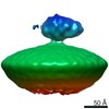


 PDBj
PDBj


 Assembly


 Mass: 79.904 Da / Num. of mol.: 25 / Source method: obtained synthetically / Formula: Br
 Mass: 18.015 Da / Num. of mol.: 195 / Source method: isolated from a natural source / Formula: H2O
 Sample preparation
 Processing