 Movie
Movie Controller

[English] 日本語
 Yorodumi
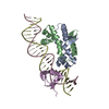
- PDB-6p0t: Crystal structure of ternary DNA complex "FX(1-2)-1Xis" containin... -
+ Open data

- Basic information
| Entry | Database: PDB / ID: 6p0t | ||||||
|---|---|---|---|---|---|---|---|
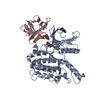
| Title | Crystal structure of ternary DNA complex "FX(1-2)-1Xis" containing E. coli Fis and phage lambda Xis | ||||||
 Components Components |
| ||||||
 Keywords Keywords | DNA BINDING PROTEIN/DNA / Protein-DNA ternary complex / DNA shape / cooperative binding / DNA BINDING PROTEIN / DNA BINDING PROTEIN-DNA complex | ||||||
| Function / homology |  Function and homology information Function and homology informationinvertasome / positive regulation of DNA recombination / sequence-specific DNA binding, bending / provirus excision / nucleoid / DNA-binding transcription repressor activity / DNA-binding transcription activator activity / chromosome organization / core promoter sequence-specific DNA binding / response to radiation ...invertasome / positive regulation of DNA recombination / sequence-specific DNA binding, bending / provirus excision / nucleoid / DNA-binding transcription repressor activity / DNA-binding transcription activator activity / chromosome organization / core promoter sequence-specific DNA binding / response to radiation / protein-DNA complex / nucleosome / DNA recombination / sequence-specific DNA binding / transcription cis-regulatory region binding / DNA-templated transcription / regulation of DNA-templated transcription / protein homodimerization activity / DNA binding / identical protein binding / cytosol Similarity search - Function | ||||||
| Biological species |   Escherichia phage lambda (virus) Escherichia phage lambda (virus) | ||||||
| Method |  X-RAY DIFFRACTION / SYNCHROTRON / MOLECULAR REPLACEMENT / molecular replacement / Resolution: 3.603 Å X-RAY DIFFRACTION / SYNCHROTRON / MOLECULAR REPLACEMENT / molecular replacement / Resolution: 3.603 Å | ||||||
 Authors Authors | Hancock, S.P. / Cascio, D. / Johnson, R.C. | ||||||
| Funding support |  United States, 1items United States, 1items
| ||||||
 Citation Citation | Journal: Nucleic Acids Res. / Year: 2019 Title: Cooperative DNA binding by proteins through DNA shape complementarity. Authors: Hancock, S.P. / Cascio, D. / Johnson, R.C. | ||||||
| History |
|
- Structure visualization
| Structure viewer | Molecule: MolmilJmol/JSmol |
|---|
- Downloads & links
-Download
| PDBx/mmCIF format | 6p0t.cif.gz | 161.9 KB | Display | PDBx/mmCIF format |
|---|---|---|---|---|
| PDB format | pdb6p0t.ent.gz | 123.4 KB | Display | PDB format |
| PDBx/mmJSON format | 6p0t.json.gz | Tree view | PDBx/mmJSON format | |
| Others |  Other downloads Other downloads |
-Validation report
| Arichive directory | https://data.pdbj.org/pub/pdb/validation_reports/p0/6p0tftp://data.pdbj.org/pub/pdb/validation_reports/p0/6p0t | HTTPS FTP |
|---|
-Related structure data
| Related structure data |  6p0sC  6p0uSC S: Starting model for refinement C: citing same article ( |
|---|---|
| Similar structure data |


-Links
 PDBj
PDBj


- Assembly
| Deposited unit | 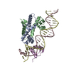
| ||||||||
|---|---|---|---|---|---|---|---|---|---|
| 1 |
| ||||||||
| Unit cell |
|
-Components
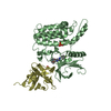
| #1: Protein | Mass: 11252.918 Da / Num. of mol.: 2 Source method: isolated from a genetically manipulated source Source: (gene. exp.) #2: DNA chain | | Mass: 8295.379 Da / Num. of mol.: 1 / Source method: obtained synthetically / Source: (synth.) #3: DNA chain | | Mass: 8291.411 Da / Num. of mol.: 1 / Source method: obtained synthetically / Source: (synth.) #4: Protein | | Mass: 6793.799 Da / Num. of mol.: 1 / Fragment: residues 1-55 Source method: isolated from a genetically manipulated source Source: (gene. exp.) Escherichia phage lambda (virus) / Gene: xis / Plasmid: pET11a / Production host: |
|---|
-Experimental details
-Experiment
| Experiment | Method: X-RAY DIFFRACTION / Number of used crystals: 1 |
|---|
- Sample preparation
| Crystal | Density Matthews: 5.23 Å3/Da / Density % sol: 76.47 % |
|---|---|
| Crystal grow | Temperature: 277 K / Method: vapor diffusion, hanging drop / pH: 7.5 / Details: 15% PEG 4000 |
-Data collection
| Diffraction | Mean temperature: 100 K / Serial crystal experiment: N | ||||||||||||||||||||||||||||||||||||||||||||||||||||||||||||||||||||||||||||||||||||||||||||||||||||||||||||||||||||||||||||||||||||||||||||||||||||||||||||||||||||||||||||||||||||||||||||||||||||||||||||||||||
|---|---|---|---|---|---|---|---|---|---|---|---|---|---|---|---|---|---|---|---|---|---|---|---|---|---|---|---|---|---|---|---|---|---|---|---|---|---|---|---|---|---|---|---|---|---|---|---|---|---|---|---|---|---|---|---|---|---|---|---|---|---|---|---|---|---|---|---|---|---|---|---|---|---|---|---|---|---|---|---|---|---|---|---|---|---|---|---|---|---|---|---|---|---|---|---|---|---|---|---|---|---|---|---|---|---|---|---|---|---|---|---|---|---|---|---|---|---|---|---|---|---|---|---|---|---|---|---|---|---|---|---|---|---|---|---|---|---|---|---|---|---|---|---|---|---|---|---|---|---|---|---|---|---|---|---|---|---|---|---|---|---|---|---|---|---|---|---|---|---|---|---|---|---|---|---|---|---|---|---|---|---|---|---|---|---|---|---|---|---|---|---|---|---|---|---|---|---|---|---|---|---|---|---|---|---|---|---|---|---|---|---|
| Diffraction source | Source: SYNCHROTRON / Site: APS / Beamline: 24-ID-C / Wavelength: 0.9793 Å | ||||||||||||||||||||||||||||||||||||||||||||||||||||||||||||||||||||||||||||||||||||||||||||||||||||||||||||||||||||||||||||||||||||||||||||||||||||||||||||||||||||||||||||||||||||||||||||||||||||||||||||||||||
| Detector | Type: DECTRIS PILATUS 6M-F / Detector: PIXEL / Date: Mar 7, 2015 | ||||||||||||||||||||||||||||||||||||||||||||||||||||||||||||||||||||||||||||||||||||||||||||||||||||||||||||||||||||||||||||||||||||||||||||||||||||||||||||||||||||||||||||||||||||||||||||||||||||||||||||||||||
| Radiation | Monochromator: Si (111) / Protocol: SINGLE WAVELENGTH / Monochromatic (M) / Laue (L): M / Scattering type: x-ray | ||||||||||||||||||||||||||||||||||||||||||||||||||||||||||||||||||||||||||||||||||||||||||||||||||||||||||||||||||||||||||||||||||||||||||||||||||||||||||||||||||||||||||||||||||||||||||||||||||||||||||||||||||
| Radiation wavelength | Wavelength: 0.9793 Å / Relative weight: 1 | ||||||||||||||||||||||||||||||||||||||||||||||||||||||||||||||||||||||||||||||||||||||||||||||||||||||||||||||||||||||||||||||||||||||||||||||||||||||||||||||||||||||||||||||||||||||||||||||||||||||||||||||||||
| Reflection | Resolution: 3.6→88.255 Å / Num. obs: 10977 / % possible obs: 99.8 % / Redundancy: 12.755 % / Biso Wilson estimate: 114.593 Å2 / CC1/2: 0.999 / Rmerge(I) obs: 0.118 / Rrim(I) all: 0.123 / Χ2: 0.936 / Net I/σ(I): 13.14 / Num. measured all: 140007 / Scaling rejects: 14 | ||||||||||||||||||||||||||||||||||||||||||||||||||||||||||||||||||||||||||||||||||||||||||||||||||||||||||||||||||||||||||||||||||||||||||||||||||||||||||||||||||||||||||||||||||||||||||||||||||||||||||||||||||
| Reflection shell | Diffraction-ID: 1
|
-Phasing
| Phasing | Method: molecular replacement | |||||||||
|---|---|---|---|---|---|---|---|---|---|---|
| Phasing MR | Model details: Phaser MODE: MR_AUTO
|
- Processing
| Software |
| ||||||||||||||||||||||||||||||||||||||||
|---|---|---|---|---|---|---|---|---|---|---|---|---|---|---|---|---|---|---|---|---|---|---|---|---|---|---|---|---|---|---|---|---|---|---|---|---|---|---|---|---|---|
| Refinement | Method to determine structure: MOLECULAR REPLACEMENT Starting model: 6P0U Resolution: 3.603→88.255 Å / SU ML: 0.54 / Cross valid method: THROUGHOUT / σ(F): 1.38 / Phase error: 26.8 / Stereochemistry target values: ML
| ||||||||||||||||||||||||||||||||||||||||
| Solvent computation | Shrinkage radii: 0.9 Å / VDW probe radii: 1.11 Å / Solvent model: FLAT BULK SOLVENT MODEL | ||||||||||||||||||||||||||||||||||||||||
| Displacement parameters | Biso max: 223.22 Å2 / Biso mean: 122.2536 Å2 / Biso min: 73.81 Å2 | ||||||||||||||||||||||||||||||||||||||||
| Refinement step | Cycle: final / Resolution: 3.603→88.255 Å
| ||||||||||||||||||||||||||||||||||||||||
| LS refinement shell | Refine-ID: X-RAY DIFFRACTION / Rfactor Rfree error: 0 / Total num. of bins used: 4
| ||||||||||||||||||||||||||||||||||||||||
| Refinement TLS params. | Method: refined / Origin x: 36.7742 Å / Origin y: -16.2079 Å / Origin z: -0.0044 Å
| ||||||||||||||||||||||||||||||||||||||||
| Refinement TLS group |
|
