- PDB-6n9y: Atomic structure of Non-Structural protein 1 of bluetongue virus -
+
Open data
ID or keywords:
Loading...
-
Basic information
Entry
Database: PDB / ID: 6n9y
Title
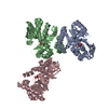
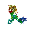
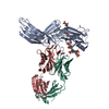
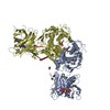
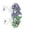
Atomic structure of Non-Structural protein 1 of bluetongue virus
Components
Non-structural protein 1
Keywords
VIRAL PROTEIN / Bluetongue Virus Non-structural protein 1
Function / homology
Orbivirus non-structural protein NS1/hydrophobic tubular protein / Orbivirus non-structural protein NS1, or hydrophobic tubular protein / Non-structural protein NS1
Function and homology information
Biological species
Bluetongue virus 23
Method
ELECTRON MICROSCOPY / helical reconstruction / cryo EM / Resolution: 4 Å
National Institutes of Health/National Institute Of Allergy and Infectious Diseases (NIH/NIAID)
AI094386
United States
Wellcome Trust
100218
United Kingdom
National Science Foundation (NSF, United States)
MCB140140
United States
Citation
Journal: Nat Microbiol / Year: 2019 Title: Atomic structure of the translation regulatory protein NS1 of bluetongue virus. Authors: Adeline Kerviel / Peng Ge / Mason Lai / Jonathan Jih / Mark Boyce / Xing Zhang / Z Hong Zhou / Polly Roy / Abstract: Bluetongue virus (BTV) non-structural protein 1 (NS1) regulates viral protein synthesis and exists as tubular and non-tubular forms in infected cells, but how tubules assemble and how protein ...Bluetongue virus (BTV) non-structural protein 1 (NS1) regulates viral protein synthesis and exists as tubular and non-tubular forms in infected cells, but how tubules assemble and how protein synthesis is regulated are unknown. Here, we report near-atomic resolution structures of two NS1 tubular forms determined by cryo-electron microscopy. The two tubular forms are different helical assemblies of the same NS1 monomer, consisting of an amino-terminal foot, a head and body domains connected to an extended carboxy-terminal arm, which wraps atop the head domain of another NS1 subunit through hydrophobic interactions. Deletion of the C terminus prevents tubule formation but not viral replication, suggesting an active non-tubular form. Two zinc-finger-like motifs are present in each NS1 monomer, and tubules are disrupted by divalent cation chelation and restored by cation addition, including Zn, suggesting a regulatory role of divalent cations in tubule formation. In vitro luciferase assays show that the NS1 non-tubular form upregulates BTV mRNA translation, whereas zinc-finger disruption decreases viral mRNA translation, tubule formation and virus replication, confirming a functional role for the zinc-fingers. Thus, the non-tubular form of NS1 is sufficient for viral protein synthesis and infectious virus replication, and the regulatory mechanism involved operates through divalent cation-dependent conversion between the non-tubular and tubular forms.
Average exposure time: 10 sec. / Electron dose: 60 e/Å2 / Detector mode: COUNTING / Film or detector model: GATAN K2 QUANTUM (4k x 4k) / Num. of grids imaged: 2 / Num. of real images: 5006
In the structure databanks used in Yorodumi, some data are registered as the other names, "COVID-19 virus" and "2019-nCoV". Here are the details of the virus and the list of structure data.
Jan 31, 2019. EMDB accession codes are about to change! (news from PDBe EMDB page)
EMDB accession codes are about to change! (news from PDBe EMDB page)
The allocation of 4 digits for EMDB accession codes will soon come to an end. Whilst these codes will remain in use, new EMDB accession codes will include an additional digit and will expand incrementally as the available range of codes is exhausted. The current 4-digit format prefixed with “EMD-” (i.e. EMD-XXXX) will advance to a 5-digit format (i.e. EMD-XXXXX), and so on. It is currently estimated that the 4-digit codes will be depleted around Spring 2019, at which point the 5-digit format will come into force.
The EM Navigator/Yorodumi systems omit the EMD- prefix.
Related info.:Q: What is EMD? / ID/Accession-code notation in Yorodumi/EM Navigator
Yorodumi is a browser for structure data from EMDB, PDB, SASBDB, etc.
This page is also the successor to EM Navigator detail page, and also detail information page/front-end page for Omokage search.
The word "yorodu" (or yorozu) is an old Japanese word meaning "ten thousand". "mi" (miru) is to see.
Related info.:EMDB / PDB / SASBDB / Comparison of 3 databanks / Yorodumi Search / Aug 31, 2016. New EM Navigator & Yorodumi / Yorodumi Papers / Jmol/JSmol / Function and homology information / Changes in new EM Navigator and Yorodumi
 Movie
Movie Controller


 Open data
 Basic information
 Components
 Keywords
 Function and homology information
 Bluetongue virus 23
 Authors
 United States,
  United Kingdom, 3items
  Citation
 Structure visualization
 Downloads & links
 Other downloads

 PDBj
PDBj Assembly
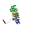
 Sample preparation
 Electron microscopy imaging
 FIELD EMISSION GUN / Accelerating voltage: 300 kV / Illumination mode: FLOOD BEAM
 Processing