 Movie
Movie Controller

[English] 日本語
 Yorodumi
- EMDB-0383: Atomic structure of Non-Structural protein 1 of bluetongue virus -
+ Open data

- Basic information
| Entry | Database: EMDB / ID: EMD-0383 | ||||||||||||
|---|---|---|---|---|---|---|---|---|---|---|---|---|---|
| Title | Atomic structure of Non-Structural protein 1 of bluetongue virus | ||||||||||||
 Map data Map data | Non-Structural protein 1 of bluetongue virus, primary map | ||||||||||||
 Sample Sample |
| ||||||||||||
 Keywords Keywords | Bluetongue Virus Non-structural protein 1 / VIRAL PROTEIN | ||||||||||||
| Function / homology | Orbivirus non-structural protein NS1/hydrophobic tubular protein / Orbivirus non-structural protein NS1, or hydrophobic tubular protein / Non-structural protein NS1 Function and homology information Function and homology information | ||||||||||||
| Biological species |  Bluetongue virus (serotype 1 / isolate South Africa) / Bluetongue virus 23 Bluetongue virus (serotype 1 / isolate South Africa) / Bluetongue virus 23 | ||||||||||||
| Method | helical reconstruction / cryo EM / Resolution: 4.0 Å | ||||||||||||
 Authors Authors | Kerviel A / Ge P | ||||||||||||
| Funding support |  United States, United States,  United Kingdom, 3 items United Kingdom, 3 items
| ||||||||||||
 Citation Citation | Journal: Nat Microbiol / Year: 2019 Title: Atomic structure of the translation regulatory protein NS1 of bluetongue virus. Authors: Adeline Kerviel / Peng Ge / Mason Lai / Jonathan Jih / Mark Boyce / Xing Zhang / Z Hong Zhou / Polly Roy /  Abstract: Bluetongue virus (BTV) non-structural protein 1 (NS1) regulates viral protein synthesis and exists as tubular and non-tubular forms in infected cells, but how tubules assemble and how protein ...Bluetongue virus (BTV) non-structural protein 1 (NS1) regulates viral protein synthesis and exists as tubular and non-tubular forms in infected cells, but how tubules assemble and how protein synthesis is regulated are unknown. Here, we report near-atomic resolution structures of two NS1 tubular forms determined by cryo-electron microscopy. The two tubular forms are different helical assemblies of the same NS1 monomer, consisting of an amino-terminal foot, a head and body domains connected to an extended carboxy-terminal arm, which wraps atop the head domain of another NS1 subunit through hydrophobic interactions. Deletion of the C terminus prevents tubule formation but not viral replication, suggesting an active non-tubular form. Two zinc-finger-like motifs are present in each NS1 monomer, and tubules are disrupted by divalent cation chelation and restored by cation addition, including Zn, suggesting a regulatory role of divalent cations in tubule formation. In vitro luciferase assays show that the NS1 non-tubular form upregulates BTV mRNA translation, whereas zinc-finger disruption decreases viral mRNA translation, tubule formation and virus replication, confirming a functional role for the zinc-fingers. Thus, the non-tubular form of NS1 is sufficient for viral protein synthesis and infectious virus replication, and the regulatory mechanism involved operates through divalent cation-dependent conversion between the non-tubular and tubular forms. | ||||||||||||
| History |
|
- Structure visualization
| Movie |
Movie viewer |
|---|---|
| Structure viewer | EM map: SurfViewMolmilJmol/JSmol |
| Supplemental images |

- Downloads & links
-EMDB archive
| Map data | emd_0383.map.gz | 28.3 MB | EMDB map data format | |
|---|---|---|---|---|
| Header (meta data) | emd-0383-v30.xmlemd-0383.xml | 12.6 KB 12.6 KB | Display Display | EMDB header |
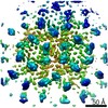
| Images | 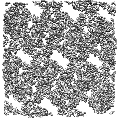 emd_0383.png emd_0383.png | 128.1 KB | ||
| Filedesc metadata | emd-0383.cif.gz | 5.8 KB | ||
| Archive directory |  http://ftp.pdbj.org/pub/emdb/structures/EMD-0383ftp://ftp.pdbj.org/pub/emdb/structures/EMD-0383 http://ftp.pdbj.org/pub/emdb/structures/EMD-0383ftp://ftp.pdbj.org/pub/emdb/structures/EMD-0383 | HTTPS FTP |
-Related structure data
| Related structure data |  6n9yMC M: atomic model generated by this map C: citing same article ( |
|---|---|
| Similar structure data |
-Links
| EMDB pages | EMDB (EBI/PDBe) / EMDataResource |
|---|
-Map
| File | Download / File: emd_0383.map.gz / Format: CCP4 / Size: 30.5 MB / Type: IMAGE STORED AS FLOATING POINT NUMBER (4 BYTES) | ||||||||||||||||||||||||||||||||||||||||||||||||||||||||||||
|---|---|---|---|---|---|---|---|---|---|---|---|---|---|---|---|---|---|---|---|---|---|---|---|---|---|---|---|---|---|---|---|---|---|---|---|---|---|---|---|---|---|---|---|---|---|---|---|---|---|---|---|---|---|---|---|---|---|---|---|---|---|
| Annotation | Non-Structural protein 1 of bluetongue virus, primary map | ||||||||||||||||||||||||||||||||||||||||||||||||||||||||||||
| Projections & slices | Image control
Images are generated by Spider. | ||||||||||||||||||||||||||||||||||||||||||||||||||||||||||||
| Voxel size | X=Y=Z: 1.07 Å | ||||||||||||||||||||||||||||||||||||||||||||||||||||||||||||
| Density |
| ||||||||||||||||||||||||||||||||||||||||||||||||||||||||||||
| Symmetry | Space group: 1 | ||||||||||||||||||||||||||||||||||||||||||||||||||||||||||||
| Details | EMDB XML:
CCP4 map header:
| ||||||||||||||||||||||||||||||||||||||||||||||||||||||||||||
 Z (Sec.)
 Y (Row.)
 X (Col.)


-Supplemental data
- Sample components
-Entire : Non-structural protein 1 (tubule)
| Entire | Name: Non-structural protein 1 (tubule) |
|---|---|
| Components |
|
-Supramolecule #1: Non-structural protein 1 (tubule)
| Supramolecule | Name: Non-structural protein 1 (tubule) / type: complex / ID: 1 / Parent: 0 / Macromolecule list: all |
|---|---|
| Source (natural) | Organism: Bluetongue virus (serotype 1 / isolate South Africa) |
-Macromolecule #1: Non-structural protein 1
| Macromolecule | Name: Non-structural protein 1 / type: protein_or_peptide / ID: 1 / Number of copies: 1 / Enantiomer: LEVO |
|---|---|
| Source (natural) | Organism: Bluetongue virus 23 |
| Molecular weight | Theoretical: 64.554984 KDa |
| Sequence | String: MERFLRKYNI SGDYANATRT FLAISPQWTC SHLKRNCLFN GMCAKQNFER AMIAATDAEE PAKAYRLVEL AKEAMYDRET VWLQCFKSF SQPYEEDIEG KMKRCGAQLL EDYRKNGMMD EAVKQSALVN SERVRLDDSL SAMPYIYVPI KEGQIVNPTF I SRYRQIAY ...String: MERFLRKYNI SGDYANATRT FLAISPQWTC SHLKRNCLFN GMCAKQNFER AMIAATDAEE PAKAYRLVEL AKEAMYDRET VWLQCFKSF SQPYEEDIEG KMKRCGAQLL EDYRKNGMMD EAVKQSALVN SERVRLDDSL SAMPYIYVPI KEGQIVNPTF I SRYRQIAY YFYSPNLADD WIDPNLFGIR GQHNQIKREI ERQVNTCPYT GYKGRVLQVM FLPIQLINFL RMDDFAKHFN RY ASMAIQQ YLRVGYAEEV RYVQQLFGKI PTGEFPLHHM MLMRRDFPTR DRSIVEARVR RSGDENWQSW LLPMIIVREG LDH QDRWEW LIDYMDRKHT CQLCYLKHSK QIPTCGVIDV RASELTGCSP FKTVKIEEHV GNNSVFETKL VRDEQIGRIG DHYY TTNCY TGAEALITTA IHIHRWIRGC GIWNDEGWRE GIFMLGRVLL RWELTKAQRS ALLRLFCFVC YGYAPRADGT IPDWN NLGS FLDTILKGPE LSEDEDERAY ATMFEMVRCI ITLCYAEKVH FAGFAAPACE SGEVINLAAR MSQMRMEY UniProtKB: Non-structural protein NS1 |
-Experimental details
-Structure determination
| Method | cryo EM |
|---|---|
 Processing Processing | helical reconstruction |
| Aggregation state | helical array |
-Sample preparation
| Buffer | pH: 7.5 Component:
| |||||||||
|---|---|---|---|---|---|---|---|---|---|---|
| Grid | Model: Quantifoil R1.2/1.3 / Material: COPPER / Pretreatment - Type: GLOW DISCHARGE / Pretreatment - Time: 30 sec. | |||||||||
| Vitrification | Cryogen name: ETHANE / Instrument: HOMEMADE PLUNGER |
- Electron microscopy
| Microscope | FEI TITAN KRIOS |
|---|---|
| Specialist optics | Energy filter - Name: GIF Quantum LS |
| Image recording | Film or detector model: GATAN K2 QUANTUM (4k x 4k) / Detector mode: COUNTING / Digitization - Frames/image: 3-19 / Number grids imaged: 2 / Number real images: 5006 / Average exposure time: 10.0 sec. / Average electron dose: 60.0 e/Å2 |
| Electron beam | Acceleration voltage: 300 kV / Electron source:  FIELD EMISSION GUN FIELD EMISSION GUN |
| Electron optics | Calibrated defocus max: 4.0 µm / Calibrated defocus min: 1.2 µm / Illumination mode: FLOOD BEAM / Imaging mode: BRIGHT FIELD / Cs: 2.7 mm |
| Sample stage | Specimen holder model: FEI TITAN KRIOS AUTOGRID HOLDER / Cooling holder cryogen: NITROGEN |
| Experimental equipment |  Model: Titan Krios / Image courtesy: FEI Company |
-Image processing
| Final reconstruction | Applied symmetry - Helical parameters - Δz: 8.83 Å Applied symmetry - Helical parameters - Δ&Phi: -17.50 ° Applied symmetry - Helical parameters - Axial symmetry: D2 (2x2 fold dihedral) Algorithm: BACK PROJECTION / Resolution.type: BY AUTHOR / Resolution: 4.0 Å / Resolution method: OTHER / Software - Name: RELION (ver. 2.0) / Number images used: 7800 |
|---|---|
| Startup model | Type of model: OTHER |
| Final angle assignment | Type: NOT APPLICABLE / Software - Name: RELION (ver. 2.0) |
-Atomic model buiding 1
| Refinement | Space: REAL / Protocol: AB INITIO MODEL |
|---|---|
| Output model | PDB-6n9y: |
